
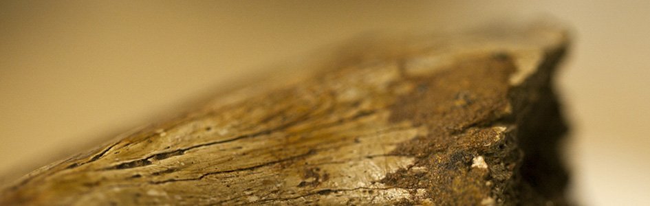
Image of a dinosaur claw by Laminin111, via Wikimedia Commons (CC BY-SA 4.0).
Dinosaur Tissue
A Biochemical Challenge to the Evolutionary Timescale
In 2005 a group of researchers, led by Dr. Mary Schweitzer, reported extracting pliable pieces of tissue from a T. rex fossil.1 Within this tissue they observed osteocytes, common cells found inside the matrix of bone. Even more surprising, they detected fragments of collagen (a common animal protein). Follow-up studies presented additional support for this discovery.2
However, the presence of tissue and protein fragments still remaining in dinosaur fossils poses a direct biochemical challenge to the standard geologic dating paradigm. If dinosaur fossils are at least 65 million years old, how has this biological material survived? How could these bones not yet be fully fossilized even after millions of years? These questions raise significant issues about contemporary dating methods.
Not surprisingly, this discovery was widely challenged. Tissue containing proteins were certainly unexpected and should not have survived millions of years of decay and fossilization. Thus, alternate ideas were offered in attempts to dismiss the tissue as “fake.” These alternatives included the suggestion that the material was from a bird carcass mixed with the fossil,3 laboratory contamination,4 and even microbial biofilm.5 While the evidence for such claims proved weak,6 it does reveal an eagerness to show the extracted material was anything other than authentic dinosaur tissue.7
Dinosaur tissue is now recognized as a “common phenomenon.”
Subsequent studies found tissue and cells in other dinosaur and reptile fossils.8 Besides collagen, proteins such as actin and myosin were also found.9 These additional discoveries helped verify the authenticity of the dinosaur tissue, and undercut the arguments of contamination. In fact, dinosaur tissue is now recognized as a “common phenomenon.”10
As additional evidence, blood vessels were carefully isolated from the femur of a duckbill dinosaur.11 They retained many physical characteristics of living animal blood vessels—pliable, translucent, and reacting to immunological based stains. The extracted vessels also contained fragments of a wide array of proteins, which is consistent with the types of proteins present in animal blood vessels.12 This work further confirms that the tissue is authentic and not biofilm or other forms of contamination.
Interestingly, despite a large body of evidence for the authenticity of the tissue, there remains a pattern of denial within the evolutionist community—presumably to downplay the ramifications of this discovery. I still receive comments from different sources (including graduate students) that they have been told the discovery was shown to be contamination. Writing for Smithsonianmag.com, Brian Switek did not even include dinosaur tissue in his 2014 list of unsolved dinosaur mysteries.13 As of 2015, Carnegie Museum of Natural History (Pittsburgh, Pennsylvania) still claimed there are “no original organic parts preserved” in fossils.14 The popular atheist website, Rationalwiki, cites outdated and misleading sources while continuing to make the claim that the tissue “has since been shown to be mistaken.”15 Add to this list all the self-appointed defenders of evolution posting commentary around the internet, scoffing that only ignorant creationists would think that a dinosaur fossil could still contain tissue, cells, and proteins.
And so it goes. Apparently many find the soft-tissue evidence much easier to dismiss than to understand and explain. Perhaps this should not be too surprising. The tissue is certainly difficult to account for within the popular geologic timescale.
Million-Year-Old Protein?
Indeed, some biological molecules, such as collagen and chitin, are chemically “tough”— resisting rapid degradation. Nonetheless, even though collagen may degrade much slower than many other biomolecules, there is no experimental evidence that collagen will survive for millions upon millions of years.16 In fact, experimental decay studies actually give an upper survival limit for bone collagen at about one million years even under ideal conditions.17 Yet evidence for collagen has been reported not only in an 85-million-year-old dinosaur fossil,18 but also in bones of a supposed 247-million-year-old reptile.19 Thus, the conundrum is evident.
In his book Dinosaur Blood,20 Dr. Fazale Rana questions the validity of these decay studies since the researchers measured degradation rates only at high temperatures. He concludes that because high temperatures will accelerate protein degradation, these studies cannot be applied to decay rates in cooler, subsurface environments.
However, Dr. Rana completely misrepresents these experiments. High temperatures are often used in protein decay studies to assure a rapid degradation, since lower temperatures can dramatically slow the rate of decay. This slower decay could extend the length of the experiment by months or even years. As long as the protein decay rate fits a predictable reaction curve,21 the Arrhenius equation can be used for converting the rate to different temperatures. Therefore, decay measured at high temperatures can be used to predict the decay rates at lower temperatures. This is a common protocol in protein biochemistry, and is well established with decades of experimental work.
Dr. Rana speculates that high temperatures may unexpectedly alter how collagen will degrade, so perhaps the Arrhenius equation cannot be properly applied. However, he fails to offer any experimental support for his conclusion. If he wants to challenge these decay studies, he needs to provide experimental evidence that collagen decay is somehow an exception to this equation. Since the decay rates are well established, he would have a distinct “uphill battle,” which might explain why he only offers conjecture as his rebuttal to the experimental work.
In addition to collagen, fragments of many other proteins have been extracted from dinosaur fossils.
Plus, as already mentioned, in addition to collagen, fragments of many other proteins have been extracted from dinosaur fossils.22 Several of these proteins (e.g., myosin, actin, and tropomyosin) are not nearly as structurally “tough” as collagen.23 In fact, studies would suggest some of these proteins degrade fairly rapidly postmortem.24 Thus, there is no experimental evidence that each of these other proteins could survive for more than just a fraction of the time that collagen can survive. Even if there were a biochemical basis that enabled collagen fragments to survive millions of years, this cannot be said about all these other dinosaur proteins.
Preservation by Iron?
By far the most popular explanation for prolonged preservation of this tissue can be referred to as the “iron model.”25 This model proposes that iron (released from hemoglobin in red blood cells) initiates reactions within the tissue that cause proteins to cross-link. By forming cross-links, proteins are potentially more resistant to enzymatic and microbial attack.
Some experimental data has been offered to support this model. Ostrich blood vessels soaked in iron solutions were preserved significantly longer than vessels soaked in water.26 At the time of this initial report, the vessels had been soaking in iron solutions for two years. While not a trivial length of time, it is very difficult to appropriately apply results of a two-year laboratory trial to the internal dynamics of subsurface fossils over a period of 68 million years.
In addition, water offers a rather poor comparison since it tends to accelerate tissue and protein degradation. Moreover, the researchers had to physically disrupt the red blood cells to achieve sufficient hemoglobin release.27 Thus, they failed to demonstrate that the iron model could even function in a natural setting.
It is also unlikely that dinosaur blood contained enough iron for this mechanism to adequately achieve preservation.28 Instead, environmental iron has been suggested as an alternate source.29 This alternative would require water to serve as the iron transporting medium, but water migrating into the fossil will accelerate tissue decay. Thus, opposing chemical dynamics would occur within the fossil (as frequently happens).30
What is more, there are several chemistry problems with the iron model. The same chemical reactions that cause cross-linking in proteins will also cause other reactions that will accelerate protein decay.31 Plus, these same chemical reactions would cause the amino acids within that protein to be chemically altered. However, numerous “intact” amino acids, such as methionine and tyrosine, are frequently found in extracted dinosaur proteins. These are highly reactive molecules that would almost certainly be chemically altered following significant iron-induced reactions within a protein molecule.32 Thus, we simply do not find the expected chemical footprint within dinosaur proteins if cross-linking were a major preservation mechanism.
Other conditions have also been proposed that might contribute to tissue preservation. However, these claims are often self-contradictory. High temperature and high/low pH can inhibit enzymatic and microbial activity, which reduces their degradative effects on tissue. Yet these temperature and pH conditions will also accelerate decay of the tissue and protein. Excluding oxygen would appear to be a significant preservation factor (oxygen will often accelerate chemical reactions). However, recent experimental evidence indicates that oxygen can help in preservation (at least for short periods of time).33 Water-free environments would certainly help preserve the tissue, but water is needed for those fossilization processes that would facilitate tissue preservation. Modest amounts of water will also help stabilize collagen.34 Furthermore, no preservation condition would protect the tissue from the devastating effect of millions of years of exposure to ground radiation.35
The “Either/Or”
Dr. Schweitzer, who continues to be one of the leading researchers in dinosaur tissue, has provided a valuable summary of the discovery. She concludes that we have “two alternatives for interpretation: either the dinosaurs aren’t as old as we think they are, or maybe we don’t know exactly how these things get preserved.”36 Fair enough. The problem is that the evolutionary community does not really consider the first alternative as a possibility. Thus, it really is not an “either/or” option. In their view the fossils must be old, therefore the tissue must somehow have survived (biochemical contradictions not withstanding).
In his “letter to creationists” Scott Buchanan states that simply because
scientists are unable at present to give a complete account of the mechanism and trajectory of the preservation of modified proteins in the dinosaur bone pores is not some unique, embarrassing case. This situation arises constantly in the course of scientific discovery.37
It is true that mechanisms can often be one of the more complex aspects to elucidate. However, Mr. Buchanan misses the point. This is not a situation where a phenomenon is readily observed, but the mechanism remains unresolved.38 Rather, no one has ever observed multi-millions of years of animal tissue preservation. The only reason there is even a quest for an unknown preservation mechanism is because evolutionary assumptions require dinosaur fossils to be at least 65 million years old.39 Remove those assumptions and there is no quest.
In point of fact, known protein decay processes actually contradict claims of 200, 100, or even 70 million years of preservation. The experimental evidence simply does not support the notion that any protein could last so long inside a fossil—let alone a variety of different proteins. Any proposed exceptions to the experimental data are simply conjecture.
What is more, with detection of flexible tissue in a supposed 550-million-year-old beard worm40 and evidence of trace amounts of protein fragments still retained in a 417-million-year-old arthropod,41 there comes a point where no amount of conjecture, inference, or proposed mechanisms (no matter how fanciful) can really even . . . well, you get the point. Buchanan chides biblical creationists for what he considers their dismissal of scientific evidence, making “them, and their version of the Christian faith, look silly.”42 At what point does the silliness turn 180 degrees?
Biblical Model
The dinosaur tissue was unexpected and is difficult to explain within the popular-held evolutionary timescale. In contrast, the tissue clearly fits within the expectations of a young earth, global flood framework. By causing a rapid, watery burial, the conditions of the Genesis Flood would enhance fossilization of dinosaurs and other creatures, thereby potentially increasing tissue survival. Protein decay data, while contradictory to a multi-million year preservation period, precisely fits within a few-thousand-year time frame. All this is very consistent with an earth that is six to ten thousand years old.
Footnotes
- Mary H. Schweitzer et al., “Soft-Tissue Vessels and Cellular Preservation in Tyrannosaurus rex ,” Science 307, no. 5717 (2005): 1952–1955, doi:10.1126/science.1108397.
- John M. Asara et al., “Protein Sequences from Mastodon and Tyrannosaurus rex Revealed by Mass Spectrometry,” Science 316, no. 5822 (2007): 280–285, doi:10.1126/science.1137614; and Mary Higby Schweitzer et al., “Analyses of Soft Tissue from Tyrannosaurus rex Suggest the Presence of Protein,” Science 316, no. 5822 (2007): 277–280, doi:10.1126/science.1138709.
- Marshall Bern, Brett S. Phinney, and David Goldberg, “Reanalysis of Tyrannosaurus rex Mass Spectra,” Journal of Proteome Research 8, no. 9 (2009): 4328–4332, doi:10.1021/pr900349r.
- Ibid.
- Thomas G. Kaye, Gary Gaugler, and Zbigniew Sawlowicz, “Dinosaurian Soft Tissues Interpreted as Bacterial Biofilms,” PLoS One 3, no. 7 (2008): e2808, doi:10.1371/journal.pone.0002808
- Mary Higby Schweitzer et al., “Molecular Analyses of Dinosaur Osteocytes Support the Presence of Endogenous Molecules,” Bone 52, no. 1 (2013): 414–423, doi:10.1016/j.bone.2012.10.010; and Mary Higby Schweitzer, Alison E. Moyer, and Wenxia Zheng, “Testing the Hypothesis of Biofilm as a Source for Soft Tissue and Cell-Like Structures Preserved in Dinosaur Bone,” PloS One 11, no. 2 (2016): e0150238, doi:10.1371/journal.pone.0151143; and Kevin Anderson, “Dinosaur Tissue or Bacterial Biofilm? Creation Research Society Quarterly 51, no. 4(2015): 259–267.
- Kevin Anderson, Echoes of the Jurassic, Chino Valley, AZ: CRS Books, 2016; and Brian Thomas, “Original Biomaterials in Fossils,” Creation Research Society Quarterly 51, no. 4 (2015): 234–247.
- Mary H. Schweitzer et al., “Biomolecular Characterization and Protein Sequences of the Campanian Hadrosaur B. canadensis,” Science 324, no. 5927 (2009): 626–631, doi:10.1126/science.1165069; and Mark Armitage and Kevin Anderson, “Soft Sheets of Fibrillar Bone from a Fossil of the Supraorbital Horn of the Dinosaur Triceratops horridus,” Acta Histochemica 115, no. 6 (2013): 603–608, doi:10.1016/j.acthis.2013.01.001; and Dawid Surmik et al., “Spectroscopic Studies on Organic Matter from Triassic Reptile Bones, Upper Silesia, Poland,” PloS One 11, no. 3 (2016): e0151143, doi:10.1371/journal.pone.0151143; and Schweiter et al., “Molecular Analyses of Dinosaur Osteocytes.”
- James D. San Antonio et al., “Dinosaur Peptides Suggest Mechanisms of Protein Survival,” PLoS One 6, no. 6 (2011): e20381, doi:10.1371/journal.pone.0020381; and Schweiter et al., “Molecular Analyses of Dinosaur Osteocytes.”
- Sergio Bertazzo et al., “Fibres and Cellular Structures Preserved in 75-Million-Year-Old Dinosaur Specimens,” Nature Communications 6 (2015): 6, doi:10.1038/ncomm8352.
- Timothy P. Cleland et al., “Mass Spectrometry and Antibody-Based Characterization of Blood Vessels from Brachylophosaurus canadensis, ” Journal of Proteome Research 14, no. 12 (2015): 5252–5262, doi:10.1021/acs.jproteome.5b00675.
- Ibid.
- Brian Switek, “The Ten Biggest Dinosaur Mysteries We Have Yet to Solve,” Smithsonian , August 8, 2014, http://www.smithsonianmag.com/science-nature/ten-biggest-dinosaur-mysteries-we-have-yet-solve-180952297/.
- Thomas, “Original Biomaterials in Fossils,” 239.
- Rationalwiki, "Evidence against a Recent Creation," http://rationalwiki.org/wiki/Evidence_against_a_recent_creation (the site reports this page was last modified Oct. 2, 2016).
- Thomas, “Original Biomaterials in Fossils;” and Anderson, “Dinosaur Tissue or Bacterial Biofilm?”
- Mike Buckley et al., “Comment on ‘Protein Sequences from Mastodon and Tyrannosaurus rex Revealed by Mass Spectrometry,’” Science 319, no. 5859 (2008): 33, doi:10.1126/science.1147046; and Mike Buckley and Matthew James Collins, “Collagen Survival and Its Use for Species Identification in Holocene-Lower Pleistocene Bone Fragments from British Archaeological and Paleontological Sites,” Antiqua 1, no. 1 (2011): 1, doi:10.4081/antiqua.2011.e1.
- Schweitzer et al., “Biomolecular Characterization and Protein Sequences of the Campanian Hadrosaur B. Canadensis.”
- Surmik et al., “Spectroscopic Studies on Organic Matter from Triassic Reptile Bones.”
- Fazale Rana, Dinosaur Blood and the Age of the Earth (Covina, CA: RTB Press, 2016), 68–70.
- Matthew J. Collins et al. “A Basic Mathematical Simulation of the Chemical Degradation of Ancient Collagen,” Journal of Archaeological Science 22, no. 2 (1995): 175–183, doi:10.1006/jasc.1995.0019; and D. J. Millward and P. C. Bates, “Protein Degradation in Skeletal Muscle: Implications of a First Order Reaction for the Degradative Process,” Acta Biologica et Medica Germanica 40, no. 10–11 (1980): 1309–1315.
- Cleland et al., “Mass Spectrometry and Antibody-Based Characterization of Blood Vessels”; and Schweitzer et al., “Molecular Analyses of Dinosaur Osteocytes.”
- According to Schweitzer et al., “Molecular Analyses of Dinosaur Osteocytes,” 421, the association of actin with α-actin and fimbrin may “stabilize the protein after [cell] death.” However, it is strictly conjecture that such association would enable actin to survive millions of years. These researchers cite studies of cell apoptosis events to support their conclusion, but such studies have not provided consistent results regarding the rate of actin degradation during apoptosis (see Celik Kayalar et al., “Cleavage of Actin by Interleukin 1 Beta-converting Enzyme to Reverse DNase I Inhibition,” Proceedings of the National Academy of Sciences 93, no. 5 [1996]: 2234–2238; and Qizhong Song et al., “Resistance of Actin to Cleavage During Apoptosis,” Proceedings of the National Academy of Sciences 94, no. 1 [1997]: 157–162). What is more, apoptosis is a specialized cell-activated process that does not involve the same cell destruction events that occur in a postmortem state.
- René Lametsch, Peter Roepstorff, and Emøke Bendixen. “Identification of Protein Degradation During Post-Mortem Storage of Pig Meat,” Journal of Agricultural and Food Chemistry 50, no. 20 (2002): 5508–5512, doi:10.1021/jf025555n; and René Lametsch et al., “Postmortem Proteome Changes of Porcine Muscle Related to Tenderness,” Journal of Agricultural and Food Chemistry 51, no. 24 (2003): 6992–6997, doi:10.1021/jf034083p; and Pål Anders Wang et al., “Post-Mortem Degradation of Myosin Heavy Chain in Intact Fish Muscle: Effects of pH and Enzyme Inhibitors,” Food Chemistry 124, no. 3 (2011): 1090–1095, doi:10.1016/j.foodchem.2010.07.093.
- Mary H. Schweitzer et al., “A Role for Iron and Oxygen Chemistry in Preserving Soft Tissues, Cells and Molecules from Deep Time,” Proceedings of the Royal Society of London B: Biological Sciences 281, no. 1775 (2014): 20132741, doi:10.1098/rspb.2013.2741.
- Ibid.
- Ibid.
- Surmik et al., “Spectroscopic Studies on Organic Matter from Triassic Reptile Bones.”
- Ibid.
- For some examples, see Anderson, Echoes of the Jurassic.
- John M. DeMassa and Edward Boudreaux, “Dinosaur Peptide Preservation and Degradation,” Creation Research Society Quarterly 51, no. 4 (2015): 268–285.
- Ibid.
- Schweitzer et al., “A Role for Iron and Oxygen Chemistry in Preserving Soft Tissues.”
- Christopher A. Miles and Michael Ghelashvili. “Polymer-in-a-Box Mechanism for the Thermal Stabilization of Collagen Molecules in Fibers,” Biophysical Journal 76, no. 6 (1999): 3243–3252, doi:10.1016/S0006-3495(99)77476-X.
- Anderson, Echoes of the Jurassic .
- Emily Ruppel, “Not So Dry Bones: An Interview with Mary Schweitzer, Biologos, July 21, 2014, http://biologos.org/blogs/archive/not-so-dry-bones-an-interview-with-mary-schweitzer.
- Scott Buchanan, “Dinosaur Soft Tissue,” Letters to Creationists, https://letterstocreationists.wordpress.com/dinosaur-soft-tissue/.
- As example, the effects of gravity are readily and continually observed by everyone, but the physical cause of gravity has proven much more difficult to understand.
- Evolutionists point to the “secure” dating of fossils by radiometric methods as a key reason why the tissue is irrelevant to the age of these fossils. Yet the accuracy and assumptions of radiometric dating methods have been seriously challenged by biblical creationists. As a rebuttal, Buchanan seeks to prove its accuracy by providing a list of radiometric dates for the Z-coal bed. All the dates on his list fall within a 4% variance (see Buchanan, “Dinosaur Soft Tissue”). However, this list is not nearly as impressive as he would lead us to believe, as it is highly “cherry picked” and unreferenced. Discordant radiometric dates are common and scattered throughout the literature (for example, see Anderson, Echoes of the Jurassic , 45–51; and Andrew Snelling. Earth’s Catastrophic Past, vol. 2. Dallas, TX: Institute for Creation Research, 2009). What is more, the materialistic underpinnings of the evolutionary timescale, and this timescale’s impact on contemporary geologic dating, is highly visible for those willing to do their homework (e.g., see John Reed. “Rocks Aren’t Clocks.” Powder Springs, GA: Creation Book Publishers, 2013).
- Małgorzata Moczydłowska, Frances Westall, and Frédéric Foucher, "Microstructure and Biogeochemistry of the Organically Preserved Ediacaran Metazoan Sabellidites ," Journal of Paleontology 88, no. 2 (2014): 224–239, doi:10.1666/13-003.
- George D. Cody et al., “Molecular Signature of Chitin-Protein Complex in Paleozoic Arthropods,” Geology 39, no. 3 (2011): 255–258, doi:10.1130/G31648.1.
- Buchanan, “Dinosaur Soft Tissue.”


Support the creation/gospel message by donating or getting involved!

Answers in Genesis is an apologetics ministry, dedicated to helping Christians defend their faith and proclaim the good news of Jesus Christ.
- Customer Service 800.778.3390
- © 2024 Answers in Genesis