
Preservation of Cellular Proteins in Dinosaur Fossils
Evidence demonstrates preservation of cellular proteins in dinosaur fossils.
News Source
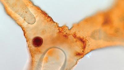
Ever since Mary Schweitzer identified red blood cells and blood vessels inside fossilized dinosaur bone in 2005, debate has raged. Could protein and cellular structures survive for millions of years? Schweitzer unveiled her latest discoveries at the 72nd annual meeting of the Society of Vertebrate Paleontology.
Schweitzer and colleagues have continued to press the case for the authenticity of preserved dinosaur soft tissue. Newly published data presented at the conference demonstrates that four kinds of protein molecules like those found in modern bone cells remain in fossils of two dinosaurs, Tyrannosaurus rex and Brachylophosaurus canadensis.
The four proteins—actin, tubulin, PHEX, and histone H4—are found in modern eukaryotes but not in bacteria. Histone H4 is a DNA binding protein, and although no DNA was sequenced, additional stains confirmed the presence of some DNA. PHEX protein is found in the osteocytes (mature bone cells) of many kinds of organisms.1 Human, rodent, and avian PHEX proteins have a high degree of homology, their amino acid sequences being very similar.2
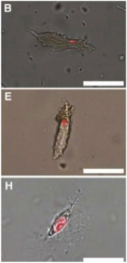
The remains of mature bone cells—osteocytes—in demineralized bone from two dinosaurs (T. rex in B, B. canadensis in E) and ostrich bone (H) are here stained with a dye for DNA, showing that at least some fragments of DNA are present in these cells. However, without being able to sequence it, its identity—suspected of course to be dinosaur in the top two photos—remains unconfirmed. Image credit: Dr. Mary Schweitzer, NC State University on Phys.Org.
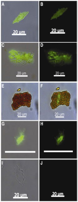
The top two rows of photos show dinosaur osteocytes outlined by fluorescing monoclonal antibody OB7.3, which targets a protein (PHEX) found only in osteocytes. (T. rex is in the first row, B. canadensis in the second.) The third row shows a dinosaur osteocyte specifically labeled by the osteocyte-specific marker while still embedded in a matrix of collagen. The fourth row shows how OB7.3 targets ostrich osteocytes too. And the fifth row shows alligator osteocytes, which are not targeted by OB7.3. Image credit: Dr. Mary Schweitzer, NC State University (Schweitzer et al. 2012), www.sciencedirect.com
Some evolutionists have insisted the purported protein-containing structures in Schweitzer’s specimens must be bacterial biofilm—a microbial slime. Biofilm proponents believe bacterial slime in old blood vessels just looks like vessels and blood cells. Even a springy texture could, they say, be produced by such “biofilm morphs.”3 Regarding these allegations, Schweitzer, showing a blank slide, said, “Here’s the data in support of a biofilm origin. We haven’t found any yet.”4
Osteocytes are encased in minerals even in living organisms. After demineralization of the fibrous collagen matrix, Schweitzer’s team used monoclonal antibodies to identify the proteins. Thus tagged, the proteins outlined star-shaped osteocytes. These results rule out “biofilm morphs” and demonstrate that cellular proteins do remain in some dinosaur fossils.5
Monoclonal antibodies must be designed in the laboratory to match specific targets. The antibody used to detect PHEX is OB7.3. It was developed in the 1980s to target chicken osteocytes.6 For this reason, much of the research performed on bone cells has been done on chickens, and it was not until the 1990s that monoclonal antibodies for mammalian osteocytes were developed.7 OB7.3 targets part of the PHEX molecule.8 And unlike the comparable mammalian antibody, OB7.3 only targets PHEX in osteocytes, not in their precursor osteoblasts.9
Schweitzer’s team used the osteocyte-specific monoclonal antibody, OB7.3, to confirm these osteocytes were really osteocytes. OB7.3 tagged PHEX in the osteocytes of ostrich bones as well as in the two dinosaurs tested, though not in alligator bone. “The PHEX finding is important because it helps to rule out sample contamination,” Schweitzer says. “Some of the antibodies that we used will react to proteins found in other vertebrate cells, but none of the antibodies react to microbes, which supports our theory that these structures are surviving osteocytes.”
Schweitzer believes additional support for the genuineness of these dinosaur proteins comes from the hypothesis that dinosaurs are “closely related to birds.”10 However, homologous proteins are only an example of common design by a common Designer, not proof of ancestry. The same can be said of many proteins that appear in homologous forms throughout the biological world. Cross-reactivity of OB7.3 with dinosaur PHEX does not demonstrate that dinosaurs evolved into birds.
The survival of protein molecules and even cellular structure in dinosaur fossils is now authenticated. The question remains, how long can such biomolecules last? It is impossible to experimentally test for millions of years of survival. Dates claiming millions of year ages for these fossils are based on unverifiable assumptions, not experimentally demonstrable proof.
These photos (see above) are showing us bone cells and DNA from dinosaurs buried around 4,300 years ago during the global Flood. We see that God, the common Designer of all creatures, naturally used the same proteins to construct the microstructure of dinosaur cells as He used for other creatures. For that reason, monoclonal antibodies developed today can detect these proteins in these ghostly osteocytes of extinct dinosaurs.
Further Reading
- #3 Soft Tissue in Fossils
- Two: Those Not-So-Dry Bones
- Collagen Coils (Collagen coils)
- DNA Decay Rate Evaluated (DNA half-life)
- More Soft Tissue in “Old” Fossils
- Persistent Protein (Persistent protein)
For More Information: Get Answers
Remember, if you see a news story that might merit some attention, let us know about it! (Note: if the story originates from the Associated Press, FOX News, MSNBC, the New York Times, or another major national media outlet, we will most likely have already heard about it.) And thanks to all of our readers who have submitted great news tips to us. If you didn’t catch all the latest News to Know, why not take a look to see what you’ve missed?
(Please note that links will take you directly to the source. Answers in Genesis is not responsible for content on the websites to which we refer. For more information, please see our Privacy Policy.)
Footnotes
- PHEX stands for PHosphate-regulating gene with homology to Endopeptidases on the X chromosome. Its name reflects the fact that the PHEX gene is expressed in mature osteocytes but not, it was thought, in their precursor osteoblasts. Recent research has shown that mammalian PHEX is also detectable in differentiated osteoblasts using a different monoclonal antibody (A. F. Ruchon et al., “Developmental expression and tissue distribution of Phex protein: Effect of the Hyp mutation and relationship to bone markers,” Journal of Bone and Mineral Research 15 (2000): 1440–1450.).
- Irene Westbroek, Karien E. De Rooij, Peter J. Nijweide “Osteocyte-Specific Monoclonal Antibody MAb OB7.3 Is Directed Against Phex Protein” Journal of Bone and Mineral Research, 17 (2002): 845–853 doi: 10.1359/jbmr.2002.17.5.845/full. Despite the similarity of mammalian and avian PHEX, monoclonal antibody OB7.3, which is made using chicken bone, does not bind to rat osteocytes. (P. J. Nijweide and R. J. P. Mulder, “Identification of osteocytes in osteoblast-like cell cultures using a monoclonal antibody specifically directed against osteocytes,” Histochemistry 84 (1986):342–347, www.springerlink.com/content/r376725052778363).
- Bacterial biofilm was found in other fossils including some from the Hell Creek Formation where Schweitzer’s original specimens were found. While bacterial biofilms are indeed found in other fossils, Schweitzer’s lab and other independent labs have repeatedly confirmed the presence of protein molecules (elastin, collagen, and hemoglobin, which are unique to vertebrates) in dinosaur fossils from other fossil sites (from “Two: Those Not-So-Dry Bones”).
- K. Wong, “Molecular Analysis Supports Controversial Claim for Dinosaur Cells,” October 18, 2012, blogs.scientificamerican.com/observations/2012/10/18/molecular-analysis-supports-controversial-claim-for-dinosaur-cells.
- Mary Higby Schweitzer, Wenxia Zheng, Timothy P. Cleland, Marshall Bern, “Molecular analyses of dinosaur osteocytes support the presence of endogenous molecules,” Bone, 2012: in press, accessed October 23, 2012, doi: 10.1016/j.bone.2012.10.010.
- P. J. Nijweide and R. J. P. Mulder, “Identification of osteocytes in osteoblast-like cell cultures using a monoclonal antibody specifically directed against osteocytes,” Histochemistry 84 (1986):342–347, www.springerlink.com/content/r376725052778363.
- One of these antibodies targets other osteocyte-related antigens, not PHEX, although PHEX is of course present in mammalian osteocytes (www.patentgenius.com/patent/6358737.html and onlinelibrary.wiley.com/doi/10.1002/jbmr.5650091104/abstract). Eventually, a mammalian antibody to PHEX was developed, but it also detects PHEX in some osteoblasts.
- Irene Westbroek, Karien E. De Rooij, Peter J. Nijweide “Osteocyte-Specific Monoclonal Antibody MAb OB7.3 Is Directed Against Phex Protein” Journal of Bone and Mineral Research, 17 (2002): 845–853, doi: 10.1359/jbmr.2002.17.5.845/full.
- A. F. Ruchon et al., “Developmental expression and tissue distribution of Phex protein: Effect of the Hyp mutation and relationship to bone markers,” Journal of Bone and Mineral Research 15 (2000): 1440–1450.
- Mary Higby Schweitzer, Wenxia Zheng, Timothy P. Cleland, Marshall Bern, “Molecular analyses of dinosaur osteocytes support the presence of endogenous molecules,” Bone, 2012: in press, accessed October 23, 2012, doi: 10.1016/j.bone.2012.10.010.


Support the creation/gospel message by donating or getting involved!

Answers in Genesis is an apologetics ministry, dedicated to helping Christians defend their faith and proclaim the good news of Jesus Christ.
- Customer Service 800.778.3390
- © 2024 Answers in Genesis