
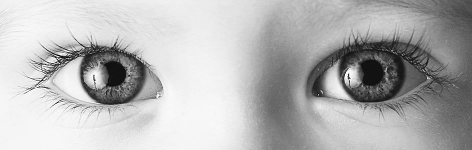
Seeing the Master Designer Through a Microbe’s Eye
News to Know
Abstract
The warnowiid’s sophisticated intracellular eye is said to have evolved from enslaved microbes.
News Sources
- National Geographic: “Single-Celled Creature has Eye Made of Domesticated Microbes”
- Cosmos: “Single Cell Eyeball Creature Startles Scientists”
- Sci-News: “Single-Celled Planktonic Organisms Have Animal-Like Eyes, Scientists Say”
- Science Daily: “Human-Like ‘Eye’ in Single-Celled Plankton: Mitochondria, Plastids Evolved Together”
Inside a single-celled organism called a warnowiid is a sophisticated structure that looks just like an eye—an ocelloid. Ocelloids look so eye-like that, when they were first discovered in the warnowiid family of plankton, some thought they were eyes of jellyfish the plankton had eaten! Scientists led by University of British Columbia’s Brian Leander have found out what ocelloids are really made of. They believe their discoveries shed light on warnowiid evolution and the evolution of eyes.
Complexity in Miniature
The ocelloid is “an amazingly complex structure for a single-celled organism to have evolved,” says lead author Greg Gavelis. “It contains a collection of sub-cellular organelles that look very much like the lens, cornea, iris and retina of multicellular eyes found in humans and other larger animals.”1 No comparable assemblage resembling a camera eye has ever been found inside any other unicellular organism. While many have eyespots—dots of photoreceptor pigment that capture light and enable a cell to orient itself in response to the light’s direction and intensity—the ocelloid is unique.
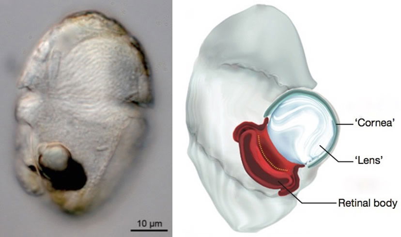
This is a warnowiid, a unicellular plankton that contains an eye-like ocelloid. The diagram shows a typical warnowiid. The ocelloid’s “lens” is cupped by a retinal body (red) and surrounded by a cornea-like envelope. Images reproduced from G. Gavelis et al., “Eye-Like Ocelloids Are Built from Different Endosymbiotically Acquired Components,” Nature 523 (July 9, 2015): 204, doi:10.1038/nature14593.
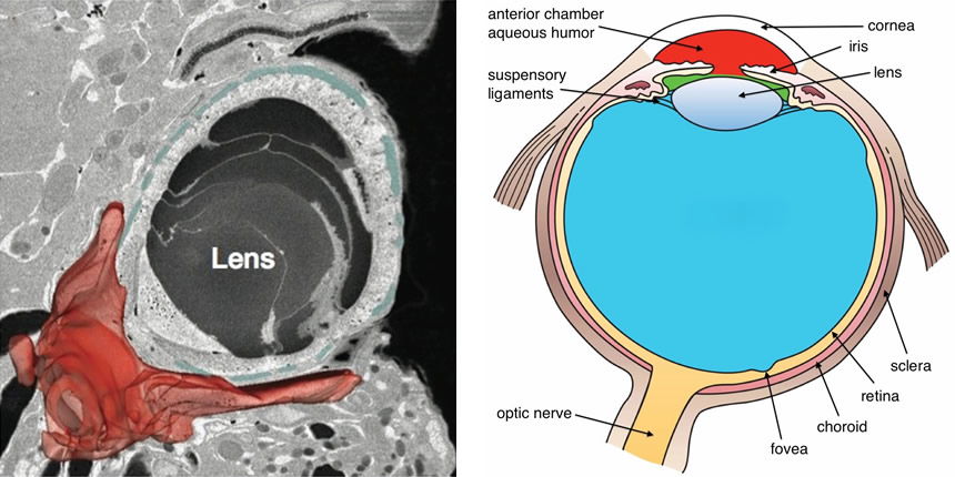
This electron micrograph of an ocelloid (left) shows how its parts compare to certain parts of a human’s “camera eye.” The ocelloid’s lens rests against a retinal body (shown in red). The lens is covered by a cornea (shown in blue). Ocelloid image reproduced from Gavelis et al., “Eye-Like Ocelloids . . . ”; human eye from Holly Fischer, Wikipedia.
Warnowiids are a family of dinoflagellates—a group of protozoans named for their whirling whip-like flagella. The many warnowiid species all have ocelloids. One even has ocelloids with multiple lenses. Warnowiids also exhibit other complex features. Some fire tiny harpoons analogous to the stinging harpoons jellyfish use to capture their dinner. Some have ballistic “pistons,” their function yet unknown.2 Some warnowiids can produce their own food through photosynthesis like plants do, but most species cannot.3 Because some warnowiids contain bits of other dinoflagellates, scientists suspect they consume their cousins. However, because warnowiids die when removed from their natural habitat, it is hard to study their behavior. Therefore, scientists have not yet seen exactly how they use their pistons and ocelloids.4
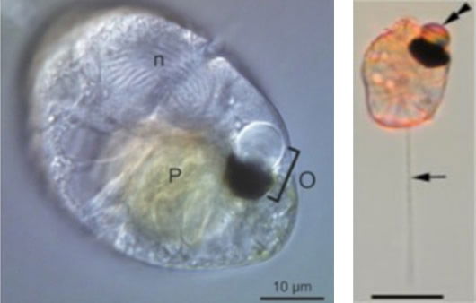
The prey (P) of the warnowiid on the left is contained in a large food vacuole. The ocelloid (O) and the nucleus (n) are also easy to see. On the right is another warnowiid with its piston extended (single arrow) and its ocelloid (double arrow) clearly visible. The dark pigmented region in each ocelloid is the retinal body. Left image from Gavelis et al., “Eye-Like Ocelloids . . .”; right image from M. Hoppenrath et al., “Molecular Phylogeny of Ocelloid-Bearing Dinoflagellated (Warnowiaceae) as Inferred from SSU and LSU rDNA Sequences,” BMC Evolutionary Biology 9 (May 25, 2009): 116, doi:10.1186/1471-2148/9/116.
Mighty Modifications
Mitochondria and chloroplasts are organelles located in the cytoplasm of many cells. Mitochondria are energy-generators. Chloroplasts normally capture light to produce food through photosynthesis. Electron microscopy revealed that ocelloids are built from these organelles.
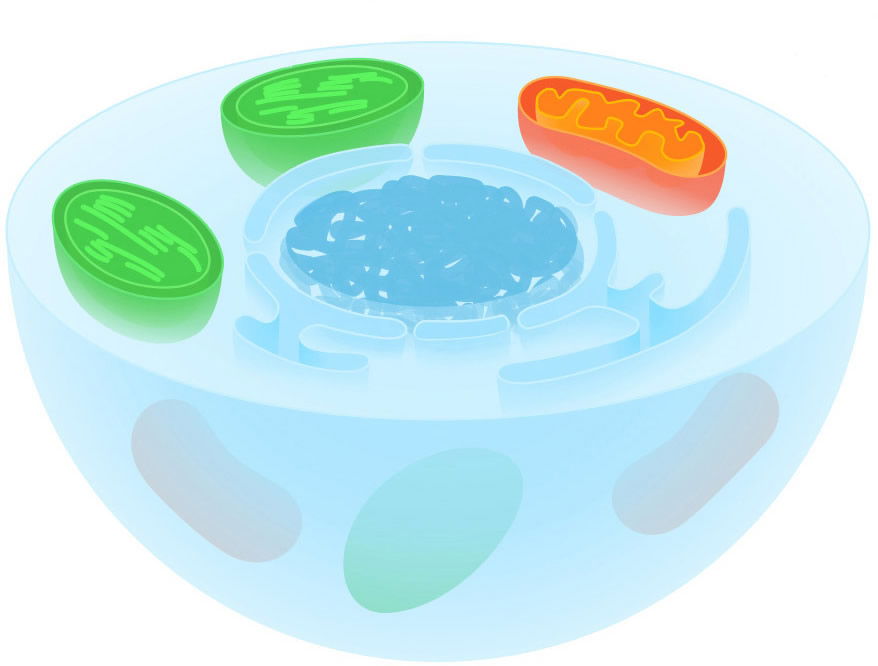
This illustration of a cell shows some important organelles. The nucleus—home to most of the cell’s DNA—is drawn in dark blue. A mitochondrion—the cell’s powerhouse—is shown in orange. Mitochondria metabolize nutrients to generate usable energy. Two chloroplasts, or plastids, are shown in green. Chloroplasts contain light-capturing pigments and enzymes that transform light energy into sugar. Unlike other cellular organelles, mitochondria and chloroplasts both contain some DNA. Image reproduced from Kevinsong, Wikipedia.
The retinal body and iris5are both made of chloroplasts. The cornea is a sheet of mitochondria. The retinal body and cornea almost completely surrounds the lens.6 Gavelis suggests the mitochondrial sheet might hold the lens in place.7 The corneal mitochondria are connected to other nearby mitochondria, and the retinal body is connected to a network of chloroplasts.

These electron micrographs of an ocelloid show that its lens (L) backs up onto a retinal body (r) and is covered by a cornea-like envelope made of mitochondria (m). Images reproduced from Gavelis et al., “Eye-Like Ocelloids . . . ”
Leander’s team found that warnowiid chloroplasts are networked into a web.8 Part of this web forms a pigment-containing9 retinal body cupping the ocelloid’s lens. They also found that the retinal body contains DNA, just as chloroplasts do. (Mitochondria and chloroplasts both contain some DNA of their own.) And even though most warnowiids are unable to perform photosynthesis, the retinal body’s DNA includes genes common in dinoflagellate chloroplasts, including some for producing light-harvesting proteins.10
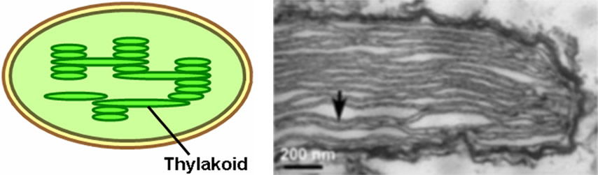
The retinal body is made of modified chloroplasts. A chloroplast, shown in the oval sketch, contains stacks of thylakoid membranes. The arrow in the electron micrograph shows ordinary thylakoid membranes in a warnowiid chloroplast that was not part of the ocelloid. Chloroplast image from Adam H., Wikipedia; electron micrograph from Gavelis et al., “Eye-Like Ocelloids . . . ”
The chloroplasts from which the retinal body is built are structurally altered. Ordinary chloroplasts are filled with stacks of thylakoid membranes, as shown above. Photosynthetic enzymes are organized on these important membranes. The retinal body is filled with stacks of membranes too. However, a retinal body’s membranes take on a wavy configuration. They also have an unusual orientation, perpendicular to the arrangement usually seen in chloroplasts. (You can see this in the pictures below.)
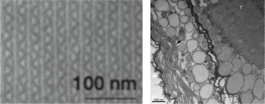
The retinal body is made of modified chloroplasts. Instead of the usual thylakoid membranes found in chloroplasts, however, the retinal body contains stacks of wavy membranes, as seen in the close-up image on the left. On the right, the waveform membranes in the retinal body (r) are easily distinguished from ordinary looking thylakoid membranes (arrow) in the ocelloid’s iris, and they are perpendicular to the usual arrangement of thylakoid membranes in a chloroplast. Images reproduced from Gavelis et al., “Eye-Like Ocelloids . . . ”
Just prior to cell division, the waveform membranes in the retinal body re-orient themselves and look just like the thylakoid membranes in ordinary chloroplasts. You can see this in the picture below, in which a retinal body’s waveform membranes are caught in the act of de-differentiating. They return to their modified wavy form in the warnowiid daughter cells.11 Thus, an ocelloid’s retinal body consists of differentiated chloroplasts.
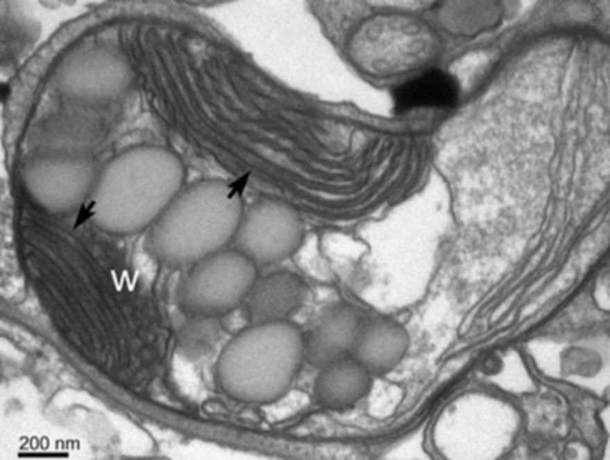
During cell division, the retinal body’s wavy membranes (w) de-differentiate and temporarily become indistinguishable from and continuous with ordinary chloroplast thylakoid membranes (arrows). This retinal body is in the midst of de-differentiating. Image reproduced from Gavelis et al., “Eye-Like Ocelloids . . . ”
Through a Microbe’s Eye
What the warnowiid can see with its ocelloid remains a mystery. With its eye-like design and genes for light-harvesting pigments, the ocelloid surely detects light, but how much more we don’t know. A single cell lacks a brain to process images, so Leander’s team does not suggest warnowiids are cognizant of what they are detecting. The retinal body’s ultrastructure, however, offers a clue. Warnowiids may pinpoint passing prey by detecting polarized light.
Remember those stacks of waveform membranes? “The internal organization of the retinal body is reminiscent of the polarizing filters on the lenses of cameras and sunglasses,” Leander explains, having “hundreds of closely packed membranes lined up in parallel.”12 So why would detecting polarized light be useful? Well, though many dinoflagellates bioluminesce, they are otherwise transparent. However, all dinoflagellates have large nuclei containing permanently condensed chromosomes. The dense nucleus polarizes light passing through the organism. Therefore, warnowiids may use their ocelloids to detect changes in light polarization produced by passing dinoflagellate prey.
Seeing the Past
Leander’s team has used cutting-edge technology to make these amazing observations. And it appears ocelloids have a visual function, though confirmation awaits study of warnowiid behavior. But can these observations provide a window into the past?
The obvious evolutionary question is whether the ocelloid camera-eye design was somehow transferred to evolving multicellular creatures and used to pattern their eyes.
The obvious evolutionary question is whether the ocelloid camera-eye design was somehow transferred to evolving multicellular creatures and used to pattern their eyes. Some scientists have proposed this. Lead author Gavelis denies this possibility, however, because he says that scenario would violate the evolutionary timeline.13 In a podcast interview he explains, “Dinoflagellates evolved far after eyes evolved in animals such as vertebrates and mollusks and jellyfish so that hypothesis doesn’t really make sense in terms of the fossil record.”14
Even though Gavelis admits the ocelloid is not the evolutionary building block for multicellular eyes, he says, “Critics of evolution often talk about how there are no transitional forms of eyes. But they’re alive and well in this case.”15
Evolutionists believe eukaryotic cells evolved by engulfing bacteria and cyanobacteria and transforming them into mitochondria and chloroplasts. Though mitochondria and chloroplasts both contain DNA of their own, they also depend on products from genes located in the nucleus. See “‘Non-evolution’ of the Appearance of Mitochondria and Plastids in Eukaryotes: Challenges to Endosymbiotic Theory” to learn more about how the dependence of these organelles on genes located in the cell’s nucleus is one of the chief arguments against this so-called “Endosymbiotic Theory.”
Leander’s team believes that warnowiids somehow united these organelles that already have an evolutionary history, modified them, and assembled them into ocelloids through additional evolutionary processes. That’s why Gavelis calls ocelloids transitional forms, even though they are a completed complex structure not apparently evolving into anything else.
Commenting on this ocelloid study, University of Exeter evolutionary biologist Thomas Richards says, “What’s so striking is they figured out how the components evolved.”16 But do these scientists really see in the ocelloid’s components its evolutionary past? Let’s take a closer look at that question.
Differentiating in the Present
What have the scientists actually seen? Leander’s team saw what ocelloids are made of. And they observed that differentiated forms of organelles normally used for metabolism and photosynthesis serve a different role as part of the complex ocelloid structure.
The warnowiid must have all the genetic information to direct such differentiation and ocelloid construction. Like cells that differentiate to perform specific functions in a multicellular organism and like stem cells in an embryo that differentiate to form each body part, organelles differentiate and assemble to form an ocelloid. But they don’t do it over millions of years. They do this every time the warnowiid reproduces, producing daughter cells just like itself, neither reenacting a primitive ancient past nor stepping up the evolutionary tree to become something new. The ocelloid is an amazing design, but it does not represent an evolutionary triumph.
Irreducibly Complex Design
So is the ocelloid a product of random evolution or a masterful design? Scientists cannot answer this question through direct observations. As evolutionists try to explain how a structure like this ocelloid evolved, they naturally look at its components and how they are assembled. Therefore, if a scientist is convinced that all life forms that exist must have evolved, then it appears obvious that evolution built the ocelloid from these organelles. The ocelloid story seems to them like a dramatic scenario, millions of years old, reenacting evolutionary steps practically before their eyes.
The apparent irreducible complexity of the ocelloid is more consistent with a biblical “God-created” worldview than with an evolutionary one.
But the issue of irreducible complexity presents a problem for the evolutionists. Without instructions, the differentiation and assembly of multiple parts into a complex whole cannot plausibly happen. All the parts of a multicomponent structure like this would need to be in place for the ocelloid to work, yet each is a specialized form of an already complex structure. What evolutionary advantage could accrue to a proto-warnowiid from the evolution of the ocelloid’s separate parts? After all, in its chloroplast-like component the thylakoid membranes are configured the wrong way for an ordinary chloroplast. The apparent irreducible complexity of the ocelloid is more consistent with a biblical “God-created” worldview than with an evolutionary one.
Converging on a Solution
Unlike ocelloids, camera eyes are ordinarily complex multicellular structures. They are found in all vertebrates and in such diverse invertebrates as scallops, squid, and jellyfish. There are also other eye designs like the crystalline eyes of chitons.17 Unable to trace the evolution of the eye through such diverse life forms, evolutionists claim that complex eyes arose through convergent evolution in many different lineages. (See the below online video Evolution: The Eyes Don’t Have It to learn more.)
Dr. Tommy Mitchell, a physician and Answers in Genesis speaker, elaborates on many problems with claims that vision evolved and shows how perfectly the human eye is designed in this online video Evolution: The Eyes Don’t Have It.
“When we see such similar structural complexity at fundamentally different levels of organization in lineages that are very distantly related, then you get a much deeper understanding of convergence,” says Leander.18 Richards, after commenting on the fact that many unicellular organisms have eyespots, writes, “The ocelloid example is striking because it demonstrates a peak in subcellular complexity achieved through repurposing multiple components.”19 That much is true, as this study demonstrates, but is that “repurposing” a function of God’s created design for these unusual organisms or the astoundingly lucky result of purposeless natural processes? Richards of course draws the expected worldview-based conclusion, claiming, “Collectively, these findings show that evolution has stumbled on similar solutions to perceiving light time and time again.”20
Convergent evolution is the idea that organisms in different lineages evolved similar features in response to similar needs. Random natural processes have no answer for how the instructions to build chloroplasts or mitochondria came to exist in the first place. It would be astounding to think that random natural processes could orchestrate ocelloid assembly—much less evolve the complex structures of mitochondria and plastids and numerous sequential enzymatic steps of their functions in the first place. This convenient term, “convergent evolution,” demands we stretch our belief in the plausibility of the implausible to a belief that the same implausible thing happened over and over again, as if a need to see were a sufficient explanation for how eyes evolved.
Our Master Designer
Indeed there is nothing particularly simple about even the simplest of our Master Designer’s creative works.
Dispensing with unverifiable evolutionary interpretations cluttering up the literature concerning warnowiids and their amazing ocelloids, we’ll simply note that they demonstrate that multicellularity is not a prerequisite for complexity. God our Creator has demonstrated His creative use of common designs by providing warnowiids with an ocelloid that structurally follows the same pattern He used in multicellular eyes but is constructed of subcellular components. Indeed there is nothing particularly simple about even the simplest of our Master Designer’s creative works.
For more information about the diversity and design of eyes in the world God made, check out these resources:
For More Information: Get Answers
Remember, if you see a news story that might merit some attention, let us know about it! (Note: if the story originates from the Associated Press, FOX News, MSNBC, the New York Times, or another major national media outlet, we will most likely have already heard about it.) And thanks to all of our readers who have submitted great news tips to us. If you didn’t catch all the latest News to Know, why not take a look to see what you’ve missed?
(Please note that links will take you directly to the source. Answers in Genesis is not responsible for content on the websites to which we refer. For more information, please see our Privacy Policy.)
Footnotes
- “Single-Celled Planktonic Organisms Have Animal-Like Eyes, Scientists Say,” July 1, 2015, http://www.sci-news.com/biology/science-warnowiid-dinoflagellate-plankton-eyes-02973.html.
- Michael Le Page, “This Single-Celled Bug Has the World’s Most Extraordinary Eye,” New Scientist, June 16, 2015, https://www.newscientist.com/article/dn27730-this-single-celled-bug-has-the-worlds-most-extraordinary-eye/.
- Loss of function is a common theme in the biology of our post-Fall world. While mutations do not create new information, they sometimes destroy existing information. Because most warnowiids cannot photosynthesize their own food even though they still have genes for some of the necessary enzymes, it is possible they have lost that ability.
- General information about warnowiids was compiled from the current study in Nature (Gavelis et al. “Eye-Like Ocelloids . . . ” doi:10.1038/nature14593) and from an additional study led by its senior author Dr. Brian Leander (Hoppenrath et al., “Molecular Phylogeny . . . ” doi:10.1186/1471-2148/9/116).
- Electron micrographs included in Gavelis’s study show that the iris-like constriction rings contain thylakoid membranes indistinguishable from those in chloroplasts.
- The “lens” is a membrane-bound structure called a hyalosome. It is broken down and synthesized anew during cell division.
- Nature podcast, July 2, 2015, http://www.nature.com/nature/podcast/index-2015-07-02.html.
- Gavelis’ team writes, “The three-dimensional reconstructions of our FIB-SEM data demonstrated that the outer membrane of the retinal body is fused to a network of adjacent plastids, forming a membranous web throughout the cell. Therefore, the retinal body appears to be a differentiated region of a larger, netlike plastid. The fact that this plastid network was not evident in previous TEM-based studies of Nematodinium suggests that hidden organelle networks could be widely overlooked in nature.” From Gavelis et al., “Eye-Like Ocelloids . . . ” doi:10.1038/nature14593.
- The retinal body of one photosynthetic species examined fluoresced bright red in green light, just as chlorophyll and some other pigments do. From Gavelis et al., “Eye-Like Ocelloids . . . ” doi:10.1038/nature14593.
- Gavelis et al., “Eye-Like Ocelloids . . . ” doi:10.1038/nature14593.
- Ibid.
- Canadian Institute for Advanced Research, “Human-Like ‘Eye’ in Single-Celled Plankton: Mitochondria, Plastids Evolved Together,” Science Daily, July 1, 2015, http://www.sciencedaily.com/releases/2015/07/150701133348.htm.
- Molecular clock dating has indicated phylogenetic markers for dinoflagellates reach back to the Precambrian, more than 570 million years ago. However, pre-Mesozoic fossils of dinoflagellates dated older than 245 million years are considered controversial. (R.A. Fensome, R.A. MacRae, and G.L. Williams, “Dinoflagellate Evolution and Diversity Through Time,” Bedford Institute of Oceanography Science Review, 1994–1995, Department of Fisheries and Oceans, Maritimes Region, pp. 45–50, http://www2.mar.dfo-mpo.gc.ca/science/review/e/pdf/dinoflagellate.pdf.)
- Nature podcast, July 2, 2015.
- Viviane Richter, “Single Cell Eyeball Creature Startles Scientists,” Cosmos, July 20, 2015, https://cosmosmagazine.com/life-sciences/single-cell-eyeball-creature-startles-scientists.
- Ibid.
-
Not all chitons have eyes. Those that do are covered with hundreds. Their lenses are made of aragonite, a form of calcium carbonate. Fossilized trilobites have calcite lenses, but aragonite lenses are unique. The West Indian fuzzy chiton is even able to respond to dark shapes rather than just changes in light intensity. Eyeless chitons, which are nevertheless covered with light sensitive cells, respond to changes in light intensity only.
Chitons supposedly took a few hundred million years to develop eyes, only acquiring them 10 million years ago, making them fairly recent additions to nature’s gallery of visual equipment. Evolutionary assumptions claim that other species evolved eyes independently during the Cambrian explosion. Why chitons evolved their eyes so “late,” why only some varieties evolved eyes when the purely light-sensitive varieties seem to survive quite nicely without them, and how they managed to acquire the genetic information to build lenses out of aragonite remain mysteries for evolutionists.
- “Single-Celled Planktonic Organisms . . . ” http://www.sci-news.com/biology/science-warnowiid-dinoflagellate-plankton-eyes-02973.html.
- T. Richards and S. Gomes, “How to Build a Microbial Eye,” Nature 523 (July 9, 2015): 166, doi:10.1038/nature14630.
- Ibid.

Support the creation/gospel message by donating or getting involved!

Answers in Genesis is an apologetics ministry, dedicated to helping Christians defend their faith and proclaim the good news of Jesus Christ.
- Customer Service 800.778.3390
- © 2024 Answers in Genesis