
The Directed Steps of Hans Christian Gram
How providence played a role in the Gram stain, which illumines the ordered pattern of bacterial cell walls and helps identify pathogens of infectious diseases
The mind of a person plans his way,
But the Lord directs his steps. (Proverbs 16:9 NASB)
This paper honors Hans Christian Joachim Gram. In published papers, he simply identified as Christian Gram. Gram devised a staining technique that is still used to identify and classify different types of bacteria. He was a Danish microbiologist, born in Copenhagen on September 13, 1853. In German microbiologist Karl Friedländer’s lab, Gram noticed that staining a smear of pneumonia bacteria with a crystal violet followed by iodine and organic solvent showed differences in various samples. “Gram-negative” bacteria have thin cell walls that allow the solvent to wash away part of the stain. “Gram-positive” bacteria appear purple in microscopy because their cell walls are thicker, so the stain cannot leave. Today, the Gram classification is fundamental to bacteria identification, classification, taxonomy, and clinical application (drug therapy) in medicine. His discovery is of great use in the identification and classification of bacteria, especially for pathogens of infectious diseases. It is also useful in deciding how to treat infections since some antibiotics are active only against gram-positive bacteria and others against gram-negative bacteria.
Insight comes from God as we move forward in biological research. He gives the seeing sight, and God illumines the mind that is open to his light. Yes, Louis Pasteur once said, “chance favors the prepared mind”—but it is providence, not just serendipity or chance, that helps the diligent man or woman make a discovery, whether that person believes it or not. These are ordered steps. God should get the glory, not just the scientists. The Master Craftsman of all things is also the Sovereign Guide who orders the steps of men. God gives insight into even seemingly accidental procedures. Previously, this was reported in the life of Alexander Fleming (Gillen and Cargill 2016; 2021). In this paper, I present the case that Christian Gram’s steps were ordered, directed, and established by God.
Introduction: Gram Stain Discovery–a Milestone in Microbiology
The cytoplasm of bacteria lacks color, and this makes it difficult for biologists to see them, especially in the tissues of people who are infected with pathogens. Therefore, microbiologists stain to impart color in them. The Gram method works by staining a sample with an iodine complex and rinsing it with a solvent. One group of bacteria retains a crystal violet-iodine complex even when rinsed by a solvent and the other group of bacteria loses the dye when rinsed. Christian Gram is reported to have serendipitously stumbled on this technique.
The Gram stain was named for Danish physician and bacteriologist Hans Christian Joachim Gram, who discovered a technique in 1883 that could differentiate all known bacteria into two categories.
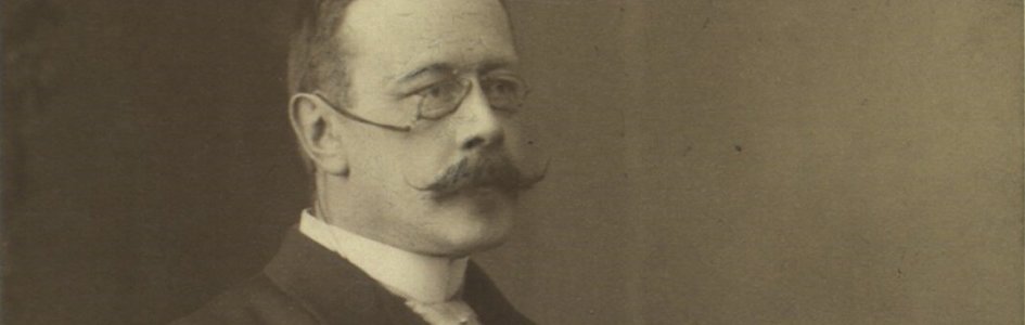
Many students of microbiology and medicine are familiar with the Gram stain, but not many know how it was discovered. The Gram stain was named for Danish physician and bacteriologist Hans Christian Joachim Gram (Fig. 1), who discovered a technique in 1883 that could differentiate all known bacteria into two categories. He would later refine the technique and publish it in 1884. The staining technique, utilizing various dyes, leaves gram-positive cells a deep purple and gram-negative cells bright red. Some may call it a serendipitous or chance event, but Dr. Gram (September 13, 1853–November 14, 1938) was a Christian, and he probably saw it as a providential event.1 He was very humble about the discovery.
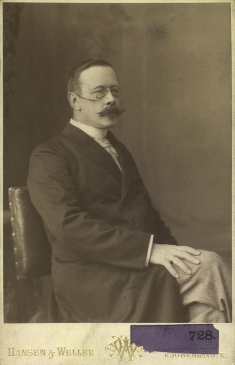
Figure 1. Portrait of Hans Christian Joachim Gram, image credit: Ww2censor via Wikipedia.
Details of the Discovery
In the 19th century, two lung diseases were the leading causes of death: tuberculosis and pneumonia. In Germany, Dr. Robert Koch had determined the cause of bacteria and a method for detecting tuberculosis; however, the causes of pneumonia were more elusive. Dr. Gram and Dr. Karl Friedländer were searching for a method that would allow visualization of cocci in tissue sections of lungs of those who had died of pneumonia. Friedländer, Gram, and their lab group were studying lobar pneumonia, working in the morgues of the City Hospital of Berlin, examining the lungs of the deceased, looking for a cause of pneumonia. Friedländer had already reported in 1882 that massive numbers of diplococci (usually with a capsule) were present in sections of lung from lobar pneumonia. These were different than the bacteria Robert Koch had found in patients with tuberculosis. Dr. Gram was familiar with both observations. Already available was a staining method designed by Robert Koch for visualizing turbercle bacilli (Gillen and Oliver 2010). Gram experimented with staining pneumococci bacteria by modifying Paul Ehrlich’s alkaline aniline solutions. Gram stained his preparations with aniline gentian violet, adding Lugol’s iodine solution for one to three minutes. When he then removed the nonspecific attributed stain with absolute alcohol, certain bacteria (pneumococci, for example) retained the color (gram-positive bacteria). Other pneumonia bacteria (Klebsiella) would remain colorless or, if counterstained with Bismarck brown, dark brown (Brock 1961).
The traditional story of Gram’s discovery is that he accidentally spilled Lugol’s iodine solution on samples that had been stained with methyl violet (crystal violet used today). He subsequently attempted to remove the iodine with a decolorizer, alcohol (today acetone-alcohol). Gram thought the iodine would ruin the stain; instead, the iodine deepened (intensified) the violet (purple). He found that in many pneumonia bacteria sections, some cells were more deeply stained purple, and some rinsed clear (pp. 81–84 Foster, 1970). The lung tissue was also free of the violet stain, allowing the bacteria to be seen against a light background. Iodine acted as a mordant for the diplococci (Streptococcus pneumonia, Fig. 2). However, it did not stick to the rod-shaped bacteria (non-encapsulated), causing pneumonia (Klebsiella pneumoniae, Fig. 3). Some types of bacteria would stain purple, and others did not stain in this multistep procedure. Gram counterstained with Bismarck brown and saw other cells as dark brown. Strangely, this method did not stain all bacteria (e.g., tuberculosis and leprosy bacteria) easily. A few years later, the German pathologist Carl Weigert (1845–1904) from Frankfurt refined the technique by replacing the Bismarck brown stain with Safranin, which demonstrated a difference (differential stain) between the cells that had a thick cell wall (later called gram-positive) and those had a thin wall (gram-negative). Gram was only 31 years old when he made his serendipitous stain.
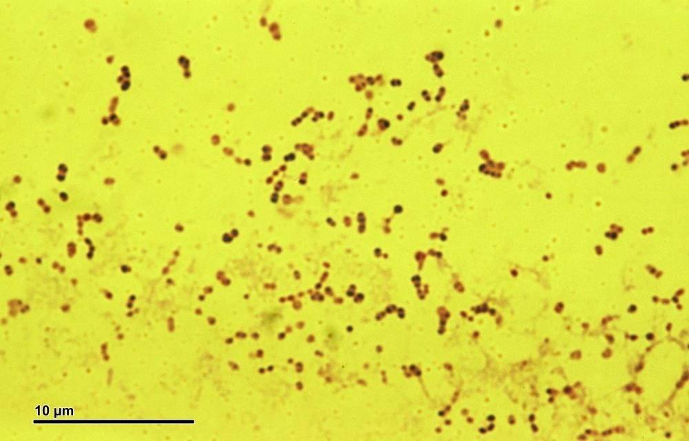
Figure 2. Streptococcus pneumonia, image credit: Petr Reischig via Wikimedia Commons.
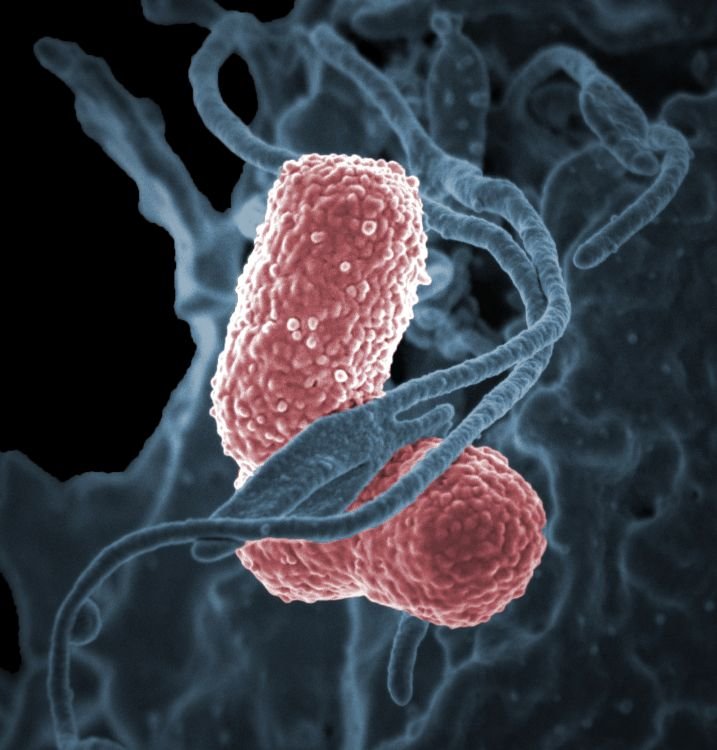
Figure 3. Klebsiella pneumoniae, image credit: Daniel Mietchen via Wikimedia Commons.
Therefore, the Gram stain is fundamental in the initial identification of bacteria. Even the bacterial cell wall itself shows further evidence of design, interwoven complexity, intricacy, and order made by the Master Craftsman.
We now recognize that this important technique identifies two large groups of bacteria: gram-positive and gram-negative. The stain reflects fundamental differences in structure and chemistry of the cell wall of the two groups (Fig. 4). Therefore, the Gram stain is fundamental in the initial identification of bacteria. Even the bacterial cell wall itself shows further evidence of design, interwoven complexity, intricacy, and order made by the Master Craftsman.
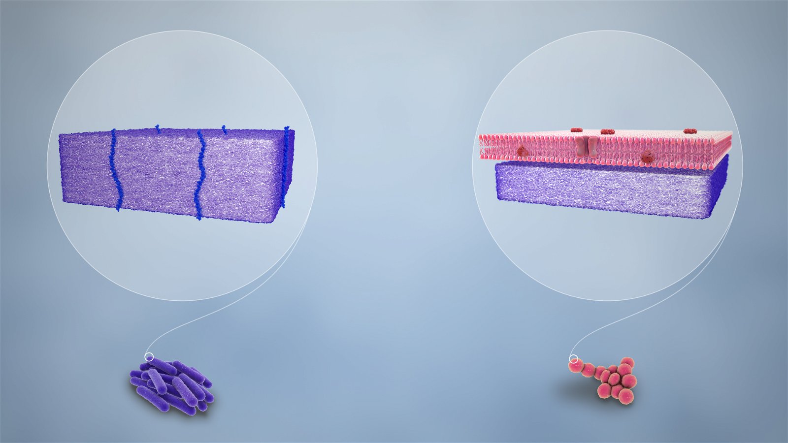
Figure 4. Purple-stained gram-positive (left) and pink-stained gram-negative (right), image credit: Manu5 via Wikimedia Commons.
Cell Walls Have Two Different Distinct Interwoven Designs
What makes some cells retain crystal violet (gram-positive) while others readily release the stain when alcohol is added (gram-negative)? Electron microscopy revealed differences in the fine structure of cell walls in gram-negative and gram-positive bacteria (Fig. 4). Gram-negative walls are not continuously associated with the underlying cytoplasmic membrane, have a thick layer of peptidoglycan, and usually are multi-layered. In gram-positive bacteria, the walls are usually closely associated with the cytoplasmic membrane and can consist of a single, amorphous layer or several layers—the structure varying in different species (Gillen 2020). The cell walls of the two categories have distinct interwoven designs, much like different woven fabrics. The distinction of these two categories has significant biological importance (classification, taxonomy) and medical application (diagnosis and treatment).
Through investigating the mechanism of antibiotic action, great advances have been made in understanding bacterial cell-wall chemistry and its intelligent design. Both gram-positive and gram-negative cells contain peptidoglycan, which forms the backbone of the rigid cell wall. Peptidoglycan is a complex polymer containing two amino sugars, glucosamine and muramic acid (from Latin murmus, meaning “wall”), and several amino acids. The muramic acid in bacterial cell walls is unique; no other known type of cell contains this sugar (muramic acid is an amino sugar acid). These simple components join to form this complex and interwoven cross-linked chemical structure. Streptococcus pneumoniae has a thick cell wall, and Klebsiella pneumoniae has a thin cell wall; hence Gram stains distinguish between the two significant pathogens. Some gram-positive organisms also contain teichoic (Greek teichos, “wall”) acids that are important for pathogen specificity.
Other bacteria have attached carbohydrates or proteins, such as the M-protein of streptococci, that contribute to virulence. Gram-negative bacteria contain lipopolysaccharides in their cell walls that sometimes contain an endotoxin. The cell walls of Gram-negative bacteria are more complex chemically than those of Gram-positive organisms. They contain lipoproteins, lipopolysaccharides, and phospholipids, but no teichoic acid. Only four or five different amino acids are found in gram-positive cell walls. Their lipid content is minimal, but they can contain teichoic acid. Cell walls illustrate the intricacy, interwoven complexity, order, and organization created by a Master Craftsman.
The Gram stain reveals the specified complexity of the two types of intricate cell walls. Now we can understand specified complexity. When a design theorist says that a string of letters is specified, he’s saying that it fits a recognizable pattern. And when he says it’s complex, he’s saying there are so many ways the bacteria cell wall could have turned out that the chance of getting any particular outcome—much less a functional or useful one—by accident is hopelessly small.
Gram Stain for Pneumonia Patients
The Gram stain is considered the most important staining procedure (Fig. 5) in microbiology. In the early years, it helped distinguish between Streptococcus pneumoniae and Klebsiella pneumoniae. Streptococcus pneumoniae was a leading cause of death in Christian Gram’s time. S. pneumoniae is the most common cause of pneumonia. Pneumonia and infectious diseases of the lower respiratory tract are the #4 cause of death worldwide even today. It is also called pneumococcus, or diplococcus, and it affects the elderly most due to poor immunity. Many young people suffer no harmful consequences. Various pneumonia infections require different treatments, and asepsis is different for gram-positive vs. gram-negative bacteria.
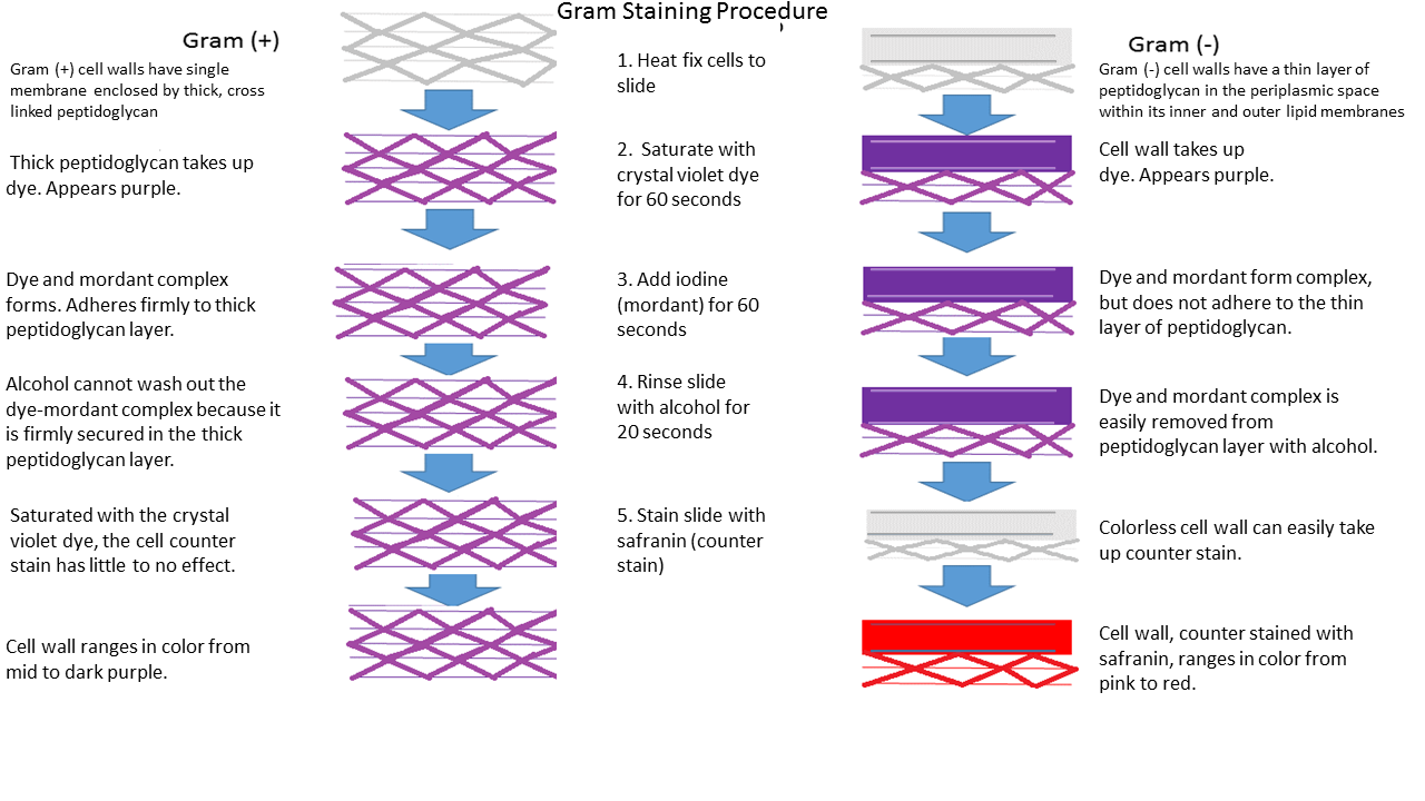
Figure 5. The gram staining procedure and its effects on gram (-) and gram (+) cell wall types, respectively, image credit: Paitynd1 via Wikimedia Commons.
The Gram stain is a planned and meaningful sequence. Changing or reversing the order of chemicals does not work.
The Gram Stain Procedure
(modified from McClintock, Gillen, and Price, 2018)
- Heat-fix and label smear.
- Flood slide with Crystal Violet for 60 seconds.
- Rinse, making sure the water stream does not go directly on the smear and wash off.
- Add Gram’s Iodine to the smear for 60 seconds. Carefully rinse with water.
- Add decolorizer to the smear for 15 seconds. (If you do not decolorize the cells long enough, the cells will stain purple regardless of the Gram stain property of the cells. If the cells are decolorized too long, the cells will remain pink regardless of the Gram stain property of the cells.)
- Rinse with water.
- Finally, add Safranin to the smear for 60 seconds. Rinse off this counterstain using water.
- Use clean bibulous paper to pat dry the slides.
- View dried and stained cells under the microscope.
The procedure is summarized with outcomes in Table 1.
Gram’s Method: Serendipitous Stain or Providential Procedure?
Providence is the guardianship and care provided by God: The Father, Jesus, and Holy Spirit. God takes care of mankind, providing insight, provision for future needs, even in medicine or clinical diagnosis. God has directed the steps of those whose discoveries have benefitted mankind.2 For other examples, look at the case of Alexander Fleming in discovering lysozymes and penicillin, Louis Pasteur in seeing the effect of attenuation of pathogens on vaccines, and Carl Fliersman in his discovery of the bacteria that caused Legionnaire’s disease. In a similar way, the providence of God was evident in how the Gram stain was discovered and refined to be of benefit to mankind. Like Louis Pasteur and Alexander Fleming, I believe his mind was prepared to see this “chance” event in a favorable light and utilize it.
Gram staining is important to human health because it helps your doctor know if you have a bacterial infection, and it determines the type of bacteria causing it. This can help in determining an effective treatment plan. Antibiotics like penicillins and cephalosporins can act upon the thick cell wall (gram-positive) made of peptidoglycan but do not work as well on thin cell walls (gram-negative). On the other hand, antibiotics like tetracycline and doxycycline act upon other cell parts and are more effective against gram-negative bacteria.
| Four Chemicals Used in Ordered Steps for Gram Stain | Color of Gram + Cells | Color of Gram – Cells |
|---|---|---|
| Primary stain: Crystal violet (30–60 sec.) | Purple | Purple |
| Mordant: Iodine (30–60) sec.) | Purple | Purple |
| Decolorizing agent: (Caution!) Alcohol-acetone (5–15 sec.) | Purple | Colorless |
| Counterstain: Safranin (30–60 sec.) | Purple | Red |
Summary
Gram staining is the definitive way to differentiate two large groups of bacteria based on their different cell wall constituents. The Gram stain procedure distinguishes between gram-positive and gram-negative groups by coloring these cells red or violet.
There is a place for prayer and recognition of God and his providence in scientific and, particularly, medical discovery.
There is a place for prayer and recognition of God and his providence in scientific and, particularly, medical discovery. The biological sciences and medicine rely on precepts provided by the concepts of biogenesis (life comes from life) and the germ theory of disease. And Joseph Lister and others who pioneered aseptic surgery ultimately profited from the pioneering work of Pasteur and his creation thinking, saving untold lives. The hand of providence was moving as Pasteur was conducting his experiments. Perhaps, R. C. Sproul summarized it best, “The invisible hand that governs the universe with ‘perfect intentionality’ has worked for the good of those who love him.”3
The cell wall is an ordered arrangement for living in different environments, and it is a marvel of design: not random but purposeful for survival and reproduction. It is beautiful to view under the microscope: purple and pink. We can thank God through Hans Christian Gram, who discovered providentially the procedure to illuminate this intricate and interwoven design to differentiate the two basic types of bacterial cell walls, providentially aiding greatly the mitigation of suffering introduced via sin and its effects (Genesis 3). The Gram stain provides critical clues for which antibiotic treatment to best use when bacteria threaten life. This has the “fingerprints” of the Great Physician.
Let the name of God be blessed forever and ever,
For wisdom and power belong to Him.
It is He who changes the times and the epochs;
He removes kings and establishes kings;
He gives wisdom to wise men
And knowledge to men of understanding. (Daniel 2:21–22 NASB)
References
Brock, T. D. Milestones in Microbiology. Englewood Cliffs, NJ: Prentice-Hall, 1961.
Foster, W. D. A History of Medical Bacteriology and Immunology. London: Heinemann Medical, 1970.
Gillen, A. L. and Oliver, J. D. “Creation and the Germ Theory: How a Biblical Worldview Helped Shape the View that Germs Make Us Sick.” Answers in Depth 4 (July 29, 2009). https://answersingenesis.org/biology/microbiology/creation-and-the-germ-theory/.
Gillen, A. L. and Oliver, J. D. “Dr. Koch, Creation, and the Constancy of Germs.” Answers In-Depth 5 (April 7, 2010). https://answersingenesis.org/biology/microbiology/robert-koch-creation-and-the-specificity-of-germs/.
Gillen, A. L. and Cargill, M. “The Signature of God in Medicine and Microbiology.” Answers in Depth 11 (March 16, 2016). https://answersingenesis.org/intelligent-design/signature-god-medicine-and-microbiology/.
Gillen, A. L. The Genesis of Germs: Disease and the Coming Plagues in a Fallen World, rev. ed. Green Forest, AR: Master Books, 2020.
McClintock, J. T., Gillen, A., and Price, M. Microbiology Lab Manual, 3rd edition. Edgewood, MD: Academx. 2018.
Sproul, R. C. The Invisible Hand. Phillipsburg, NJ: Presbyterian & Reformed Publishing. 1996.
Dr. Alan L. Gillen is a professor of biology at Liberty University.

Footnotes
- Though Hans Christian Joachim Gram was his full name, he simply identified himself as Christian Gram in published papers, signed papers, and photographs. See also “Hans Christian Gram Biography, Age, Death, Career, Achievements, Family, Wiki & More,” September 13, 2019, https://www.celebrityborn.com/biography/hans-christian-gram/16333.
Gram, C. 1884. “Ueber die isolirte Farbung der Schizomyceten in Schitt-und Trockenpreparaten,” Fortschritte der Medicin, 2: 185-189. - Proverbs 20:24 — A person’s steps are directed by the Lord. How then can anyone understand their own way? Other passages similar include Psalm 37:23.
- R. C. Sproul, The Invisible Hand (Phillipsburg, NJ: Presbyterian & Reformed Publishing, 1996), book cover.


.jpg)
.jpg)
Support the creation/gospel message by donating or getting involved!

Answers in Genesis is an apologetics ministry, dedicated to helping Christians defend their faith and proclaim the good news of Jesus Christ.
- Customer Service 800.778.3390
- © 2024 Answers in Genesis