
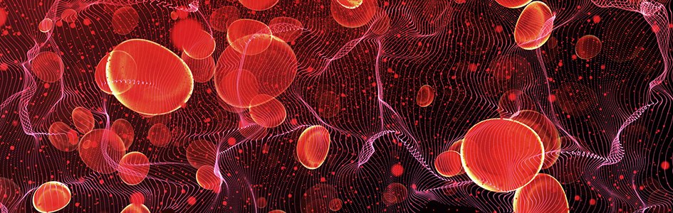
Life Is in the Blood
Keywords: Blood, Circulation, Leeuwenhoek, red blood cells, white blood cells, capillary, life is in the blood, erythrocyte, leukocyte, rouleaux
Abstract
It takes about 60 seconds for all the blood in your body to complete its journey. It travels from your heart to your extremities and returns, there and back again. Blood moves with the rapid current of the great arterial rivers and through the smallest capillary creeks. William Harvey first noticed circulation (1628) through the heart into arteries and veins; however, he could not see how they connected since he did not have a microscope. The man who first described this was Anton van Leeuwenhoek about 46 years later (1674). Then, J. J. Lister and Thomas Hodgkin described the rouleaux formation or stacking of RBCs through a capillary bed. All of these men mentioned above were committed Christians.
Three hundred years ago (1719), Leeuwenhoek was providing his most detailed account of red corpuscles and capillary circulation. He also provided an accurate measurement of 0.003 inches (actual: 6.2–8.2 µm) for human blood cells and described many different types in animal cells. This was about 45 years after he first described them as a young man (1674).
The human body produces two million blood cells deep in its bone marrow per second and pumps 1500–2000 gallons per day. Once formed, those red blood cells (RBCs) move into the bloodstream with white blood cells and platelets, all circulating through 60,000 miles of arteries, veins, and capillaries in the human body. Leeuwenhoek described the connections between arteries and veins as capillaries and demonstrated what Harvey could only postulate.
Blood reveals much about the majesty of our Creator and Master Craftsman, irreducible complexity, and the health or disease state of the human body.
Knowledge of the blood and circulatory system gives us insight into spiritual, biological, and clinical applications. Blood reveals much about the majesty of our Creator and Master Craftsman, irreducible complexity, and the health or disease state of the human body. Capillaries are the smallest blood vessels through which blood cells can move through in single file. This blood vessel network knitted with lymphatic capillaries shows an interwoven complexity, thus revealing the fearfully and wonderfully made design of our body (Gillen, 2009). In this article, we also show a biblical worldview and notable Christians who expounded the biblical concept that “Life is in the Blood.”
For the life of the flesh is in the blood. (Leviticus 17:11)
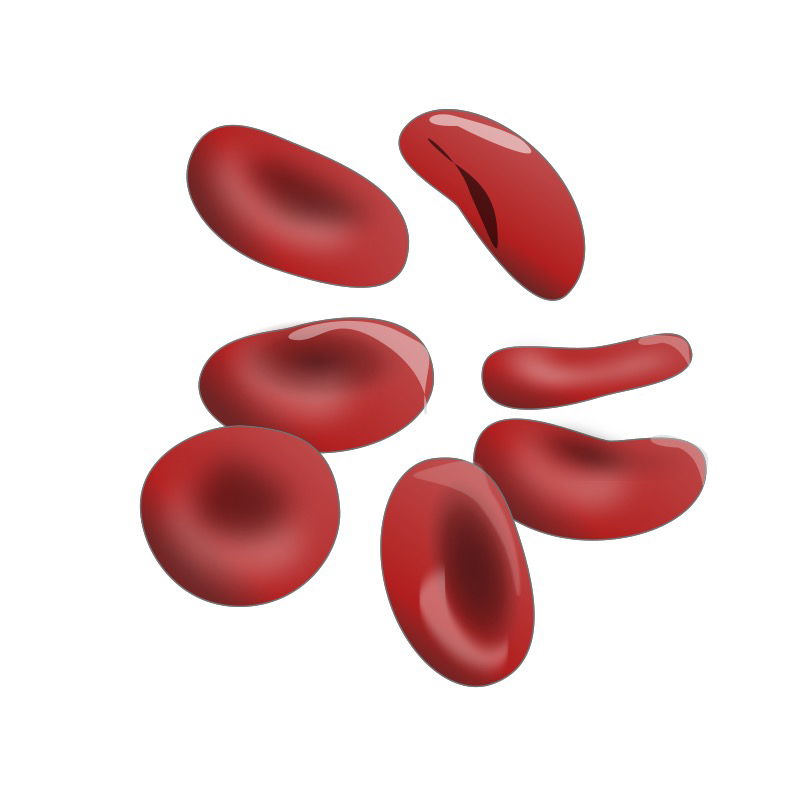
Fig. 1. Red blood cell, an erythrocyte. Notice the biconcave shape in 3D. Image by Jessica Polka via Wikimedia Commons.
Introduction
Blood Cells are the Majority
It is estimated that the body of an average man contains around 30 trillion eukaryotic cells. The majority of the cells in our bodies are actually RBCs (Fig. 1). Although they make up over 83% percent of our body in number, they constitute a lesser mass. This is because RBCs only measure on average 8 micrometers in diameter, which is 10 times smaller in diameter than an average human hair.
Experts have calculated the number of RBCs. This is achieved by taking an average blood volume of 4.9 liters (L) multiplied by a mean RBC count of 5.0x10¹² cells/L. This could be verified by looking at your routine complete blood count. Normal values range from 4.6–6.1x10¹² cells/L for males and 4.2–5.4x10¹² cells/L for females. This calculates to a total of 2.5x10¹³ RBCs. They clearly make up the overwhelming majority of our body cells (Sender, Fuchs, and Milo 2016).
Life Is in the Blood
Blood is a rich scarlet soup of proteins and cells that keeps us alive. A few nights working in an emergency room would probably convince you that the body is just a huge bag of blood. Actually, in an average 70-liter human body, only five liters, or 7% by volume, is blood. Normally, blood is found in the heart, in blood vessels, and in the sinusoids of the marrow, liver, and spleen. Of the average five liters of blood, only 2.25 L, or 45%, consists of cells. Erythrocytes, leukocytes, and platelets are the formed elements of blood. The rest is plasma that consists of 91.5% water (by weight) and 8.5% solids (mostly albumin). Of the 2.25 L of cells, only 0.037 L (1.6%) are leukocytes (Fig. 2). The entire circulating leukocyte population, if purified, would fit in a coffee cup. The total circulating platelet volume is even less, about 0.0065 L, or about one teaspoonful. Because blood is the connecting fluid to all the body systems and is the substance upon which all body cells are dependent, we explore blood as it relates to all the physiological patterns.
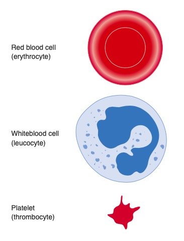
Fig. 2. Three types of blood cells: red blood cell, white blood cell, and platelets. Image by Cancer Reserach UK uploader via Wikimedia Commons.
RBC Form and Function
A study of erythrocytes or RBCs illustrates the correlation between structure and function. RBCs are structurally the simplest cells in the body. RBCs are highly specialized for their oxygen transport function. Each one contains an amazing 280 million hemoglobin molecules. Although RBCs initially have a nucleus in humans, they lose it as they mature (Fig. 3). Since RBCs do not have nuclei, all their internal space is available for oxygen transport. The shape of the RBC facilitates its function. A biconcave disc has a greater surface area for its volume than a sphere or a cube. This large surface area allows for the diffusion of gas molecules, vitamins (B12, B6, C, E, folate, riboflavin, pantothenic acid, thiamine), and amino acids. Their size and shape facilitate moving through different capillaries and maximizes oxygen delivery to all the body cells. The shape of the RBCs is clearly designed and not formed by chance. Efficient gas transport requires erythrocytes to pass through very narrow capillaries, and this constrains their size. Some erythrocytes are slightly larger than capillary vessel diameter, which are as narrow as 3 µm, and they require flexibility to move through vessels freely.
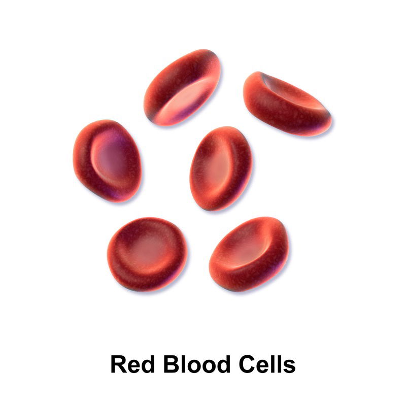
Fig. 3. Mature red blood cells, an erythrocyte. Notice the center is without a nucleus. Image by BruceBlaus via Wikimedia Commons.
Erythrocytes arise from blast precursors in the marrow over five days. Then they are released into the blood as reticulocytes, distinguishable from regular erythrocytes only with special stains. The reticulocyte changes to an erythrocyte in one day and circulates for 120 days before being destroyed in the reticuloendothelial system. The basic function of the RBC is the creation and maintenance of the body’s internal environment, providing oxygen and eliminating carbon dioxide and body waste.
Leeuwenhoek’s Legacy of Red Corpuscle Discovery
Antony van Leeuwenhoek (Fig. 4) found great joy in God’s smallest creatures and human body cells. He first discovered RBCs in humans and animals in his youth. But he was nearly 90 years old (1719) when he drew them in detail. The Dutch haberdasher retained a child-like joy of discovery from his youth until his death at age 90. He lived to see tiny cells though his homemade microscopes. He loved to grind and focus a new lens in order to see the unseen world. Leeuwenhoek spent countless hours grinding tiny lenses and looking through them. This Christian lay biologist even used candlelight to see specimens at night. For him, the amazing diversity of tiny cells and life forms revealed under his homemade microscopes glorified God as much as looking at stars through a telescope (Gillen and Oliver 2012).
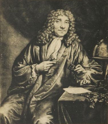
Fig. 4a. Antoni van Leeuwenhoek’s portrait. He is commonly known as the “Father of Microbiology.” Leeuwenhoek is known for his advancements with the microscope and descriptions of many bacteria and protozoa. Image by Tryphon via Wikimedia Commons.
It is to be hoped then, that the enquirers into Nature’s works, by searching deeper and deeper into her hidden mysteries, will more and more place the discoveries of the truth before eyes of all, for as to produce aversion to the errors of former times, which all those who love the truth ought diligently to aim at. For we cannot in any better manner glorify the Lord and Creator of the Universe than that, in all things, how small forever they appear to our naked eyes, which yet have received the gift of life and power if increase, we contemplate the display of his Omniscience and Perfections with the utmost admiration (Hoole, 314).
Anton van Leeuwenhoek was not the inventor of the microscope; however, he may have advanced it more than anyone else. He is well known as the first to see bacteria and protozoans. It is less known that he frequently wrote about blood and its circulation through experimentation, especially the blood flow through capillaries. Leeuwenhoek observed RBCs, measured (0.079 mm or 0.0031 inches), flowing through capillaries. Leeuwenhoek’s first written description of human red blood cells (his own) was in a letter to Constantine Huygens on April 5, 1674, and later in another letter addressed to the Secretary of the Royal Society of London on April 5, 1674. The first publication of the observation of globules in blood occurred in April 1674 in the Philosophical Transactions of the Royal Society of London in which Leeuwenhoek published his 1673 observations reported in the letter to Huygens (Hoole 1798).
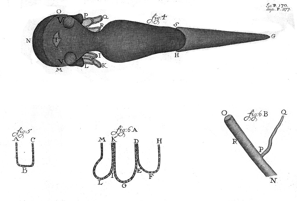
Fig. 4b. Antoni van Leeuwenhoek’s drawings of capillaries. Leeuwenhoek was the first to see and describe the circulation of blood corpuscles in capillaries. Image by Fæ via Wikimedida Commons.
In a letter dated August 14, 1675, Leeuwenhoek wrote about his remarkable discovery that “those sanguineous globules in a healthy body must be very flexible and pliant, if they are to pass through the small capillary veins and arteries, and that in their passage they change into an oval figure, reassuming their roundness when they come into a larger room.” The first known drawings of animal RBCs by Leeuwenhoek were contained in letters dated March 3, 1682 (salmon RBCs) and July 16, 1683 (frog RBCs). Leeuwenhoek’s first drawing of human RBCs (long thought to be the first drawing ever of human RBCs) was contained in a letter dated July 7, 1700 (Hoole 1798).
Rouleau Formation Reveals Disease
Many people are familiar with Anton van Leeuwenhoek and Lord Joseph Baron Lister, but few are aware of the critical contributions of Lord Lister’s father, Joseph Jackson Lister (Gillen and Oliver 2019). The senior Lister (J. J. Lister) was responsible for improving the microscope that was used in Lord Lister’s later discoveries. The improved light microscope enabled the viewing of RBCs as biconcave discs, often moving in stacks circulating through a capillary called rouleaux (Fig. 5). Rouleaux (rouleau is singular) are stacks or aggregations of RBCs that form due to the discoid shape of the cell. A rouleau occurs when the plasma protein concentration is elevated, causing an increased ESR (erythrocyte sedimentation rate). This is a non-specific indicator of the presence of disease. A rouleaux of RBCs looks like “melted” cherry Life Savers® (candy) stacked together in a roll, or a stack of coins rolled up.
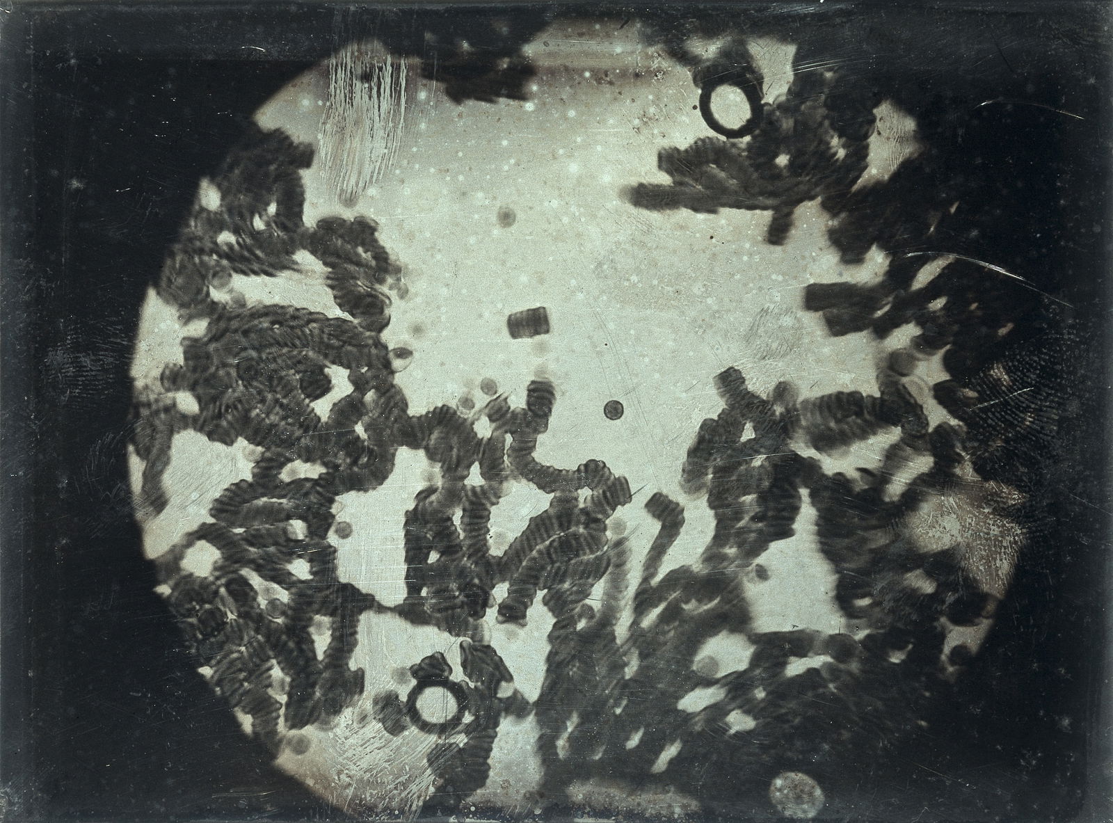
Fig. 5. Rouleaux of blood corpuscles. This is likely a microscopic view that J. J. Lister and Thomas Hodgkin saw of blood cells stacked on top of each other. Image by Fæ via Wikimedia Commons.
The main significance to J. J. Lister’s new 1826 microscope was to provide a superb quality to the viewing of microscopic images and invent a mechanism to clearly see cellular features, such as the biconcave shape and measure its exact size. In addition, J. J. Lister was a friend of Dr. Thomas Hodgkin, a fellow Quaker and physician at a prominent hospital in London. Using the “newly” invented microscopes (1826), J. J. Lister and Hodgkin conducted studies on blood and co-authored scientific papers. They were the first to describe the elegant and intelligently designed, biconcave disc-shape of healthy RBCs (erythrocytes) and their tendency to gather like a stack of dishes in a roll. Later, Dr. Thomas Hodgkin would describe a cancer of blood (lymphoma), now called Hodgkin’s disease in his honor. Rouleaux formation usually means disease. It revealed cancer in the time of J. J. Lister and Hodgkin. In our times, we also see it in certain infectious diseases and in malaria. I (ALG) have seen it personally with falciparum malaria.
Rouleaux formation can happen in cerebral malaria, but the actual RBCs infected with Plasmodium tend not to go through brain capillaries; they stay outside the brain tissue.
Lord Joseph Baron Lister (JJL’s son) continued these studies as a medical student and later studied white blood cells and their role in wound healing and inflammation in frogs and applied his learning to human surgery. It might be noted that a major part of the Listers’ success was built upon the knowledge of a proficient microscope, and Lord Lister’s knowledge of blood coagulation and inflammation were also based upon his father’s previous experience.
Cleansing Power of the Blood through Phagocytosis
One of the earliest observations regarding the immune system was the ability of the immune system to recognize “self” from “non-self.”
One of the earliest observations regarding the immune system was the ability of the immune system to recognize “self” from “non-self.” Special white blood cells called phagocytes are designed to engulf and digest invaders by phagocytosis (Figs. 6 and 7). In animal circulatory systems, phagocytes monitor tissues as they travel throughout the body. The discovery of the protective role of phagocytes was first seen in starfish and was a milestone in immunology. In 1882, Russian biologist Elie Metchnikoff collected transparent starfish larvae on a beach in Europe, pierced them with rose thorns, and waited to see what happened. A day later, he saw that phagocytes had collected at the site of injury. The thorn in a starfish larva is not unlike a splinter having pierced the skin of a person in terms of the immune system rapidly responding to an antigen. Joseph Lister would later pick up on this concept and also saw the role of white blood cells in inflammation, suppuration, and wound healing.
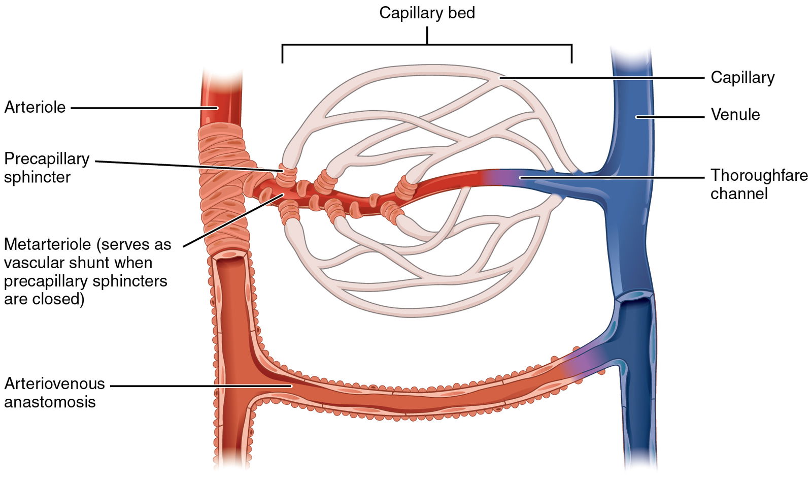
Fig. 6a. Capillary bed diagram. Capillaries are blood vessels that connect arterioles with venules by way of a capillary bed. It is here where fluid known as interstitial fluid (lymph) leaks out to supply our cells with nutrients. Image by CFCF via Wikimedia Commons.
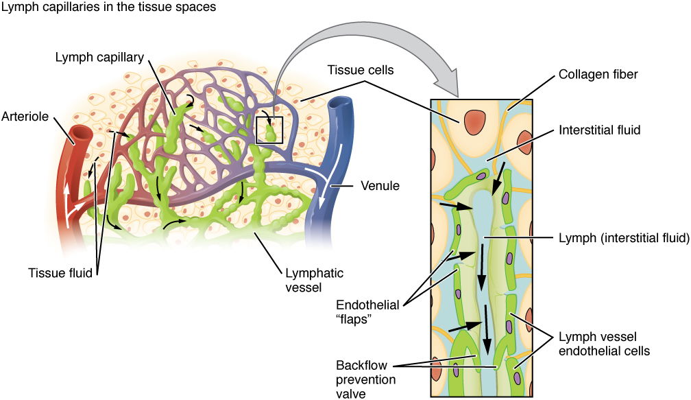
Fig. 6b. Lymphatic Capillaries. Parts and flow diagram of a lymphatic capillary. Image by CFCF via Wikimedia Commons.
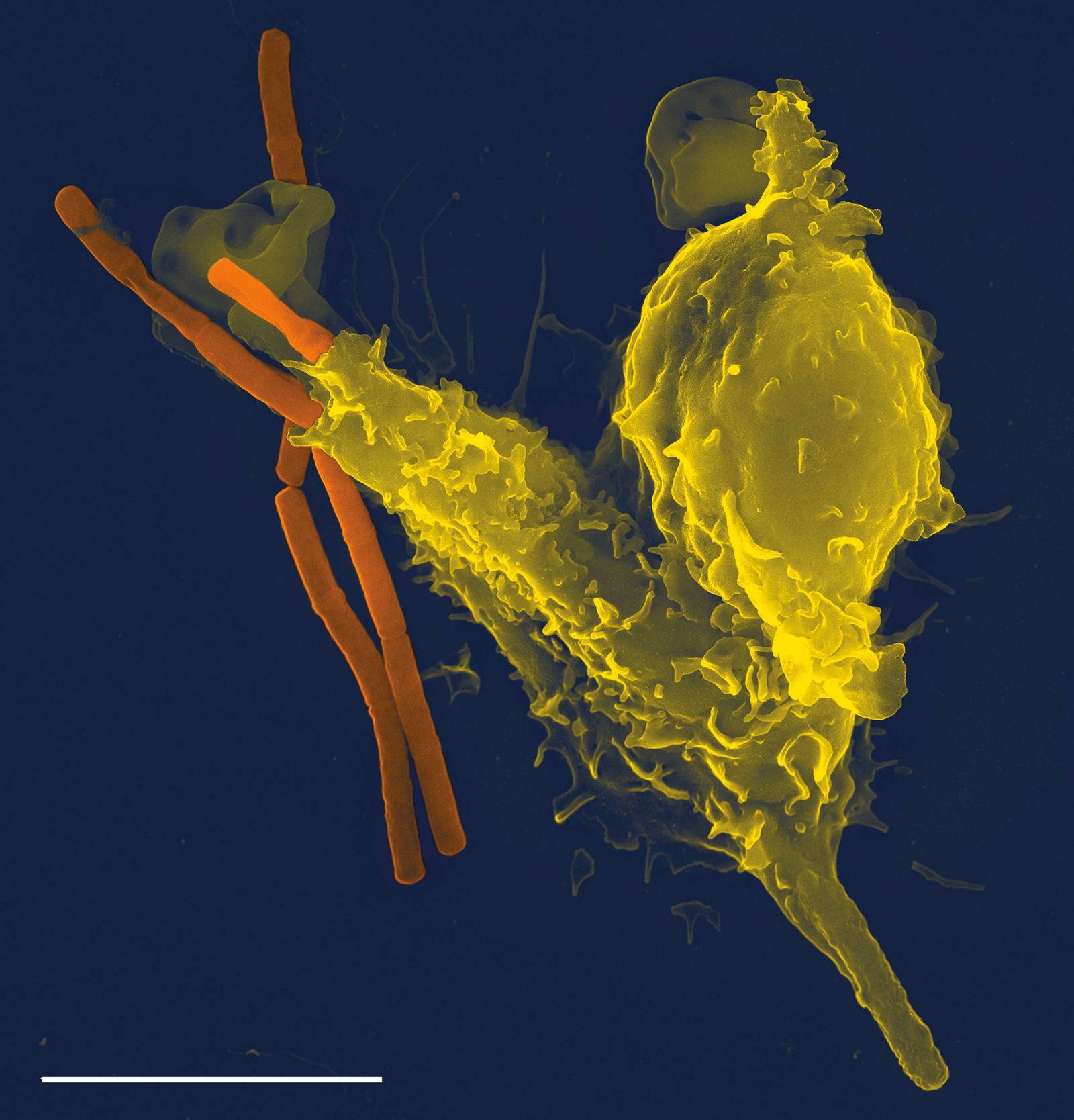
Fig. 7. Phagocytosis of a Neutrophil ”eating” anthrax bacteria. Image by TimVickers via Wikimedia Commons.
Phagocytosis is the process in which cells engulf particles in a manner similar to the way an amoeba eats (Fig. 7). The human body uses it to both cleanse itself of aged and dying cells, as well as in defense against a microbe, parasite, or toxin. Phagocytosis (Phago: eat; Cyte: cell; Osis: process) is a process of engulfing a solid particle by a phagocyte or amoeba to form an internal phagosome (Gillen and Conrad, 2014). The cell or particle becomes surrounded by cytoplasmic extensions called pseudopods that ultimately fuse. Thus, the particle becomes surrounded by a membrane derived from the plasma membrane and contained within an organelle analogous to a food vacuole in an amoeba. This vacuole then fuses with lysosomes so that the ingested particle and the digestive enzymes still are separated from the cytoplasm by a continuous membrane. The four basic phases of phagocytosis are (1) chemotaxis; (2) adherence; (3) ingestion; and (4) digestion. In reality, phagocytosis is much more complex than this and a process that Dr. Michael Behe might call irreducibly complex (Behe 1996, pp. 102–103).
Cleansing in the Lymph Capillary: An Interwoven Design
Lymphatic capillaries show interwoven complexity; we are knit together and fearfully and wonderfully made. In the circulatory and lymphatic systems, we observe parts that may be described as intertwining, interlocking, and interlacing. The interdependent parts, woven together, appear to be the “fingerprints” of a divine Craftsman. We might describe this collection of intertwining, interlocking, and interlacing parts as interwoven complexity (Gillen 2009).
Some of the most perceptible pieces of evidence of a skilled craftsman include high interdependence and harmonious working of diverse body structures and systems.
For you formed my inward parts; you knitted me together in my mother’s womb. I praise you, for I am fearfully and wonderfully made (Psalm 139:13–14).
The fabric-of-life idea reveals that the Creator purposely wove together both blood and lymphatic capillaries.
The fabric-of-life idea reveals that the Creator purposely wove together both blood and lymphatic capillaries. The Master Weaver also stitched a network of vessels containing dozens of specialized cells to form a finished masterpiece fabric that surpasses all others in intricacy, specificity, and beauty. Upon closer examination, one can see that no matter what level of the body is analyzed (e.g., organ, tissue, cellular, or molecular), elegant design is seen.
The lymphatic capillary system is awesome and distinct in nature. It is found throughout the body but is most common in places like lymph nodes. The design of the interwoven lymph nodes is further evidence of a skilled Weaver, or Master Craftsman. It is knit together with the circulatory system. The transport of the lymph begins in microscopic “blind-ended” lymphatic capillaries. The capillaries weave between the tissue’s cells and blood capillaries in the loose connective tissues of the body. Lymphatic capillaries are widespread throughout the body and are now known in limited elongations of the meninges of the brain. Although similar to blood capillaries, lymphatic capillaries are so remarkably permeable that they were once thought to be open at one end like a straw. We now know they owe their permeability to the two unique structural modifications: the endothelial cells form walls of lymphatic capillaries that are not tightly joined. Instead, the adjacent cells overlap each other loosely forming easily opened flap-like mini-valves. Collagen filaments attach the endothelial cells to surround structures so that any increase in interstitial fluid volume opens to the mini-valves rather than causing them to collapse (Marieb and Hoehn 2020). Lymphatic capillaries filter lymph and indirectly “wash” the blood of waste and pathogens.
Anyone looking at this rich tapestry would immediately praise the body of its intricate and complex design. Yet the very same people will claim that the complex human body happened by chance.
Blood and lymph capillaries cleanse the body’s fluids of particles, pathogens, and parasites. Macrophages, neutrophils, and dendritic cells filter the blood and lymph. Anyone looking at this rich tapestry would immediately praise the body of its intricate and complex design. Yet the very same people will claim that the complex human body happened by chance. A clear demonstration of the cleansing design of the blood and lymph can be seen by observing pus under a microscope, revealing many neutrophils.
The Revealing Power of the Blood
Blood is a river of life that surges within us, and a blood test reveals how healthy we are. Blood has enormous importance in the practice of medicine. Clinicians examine it more often than any other tissue trying to determine the cause of a disease in their patients. In my microbiology and parasitology classes, we talk about its revealing power in interpreting a complete blood count and how abnormal counts reveal an infectious or parasitic disease (Table 1).
Table 1. The revealing power of the blood.
- Bacteria infection – Neutrophil and/or macrophage count high
- Viral infection – Lymphocyte count high
- Toxoplasmosis infection – Lymphocyte count high
- Malaria infection – “signet ring” inside RBCs
- Worm parasite infection – High eosinophil count
- Ectoparasites (ticks) – High numbers of basophils
- Mononucleosis –High numbers of enlarged B-lymphocytes that resemble monocytes caused by Epstein Barr virus
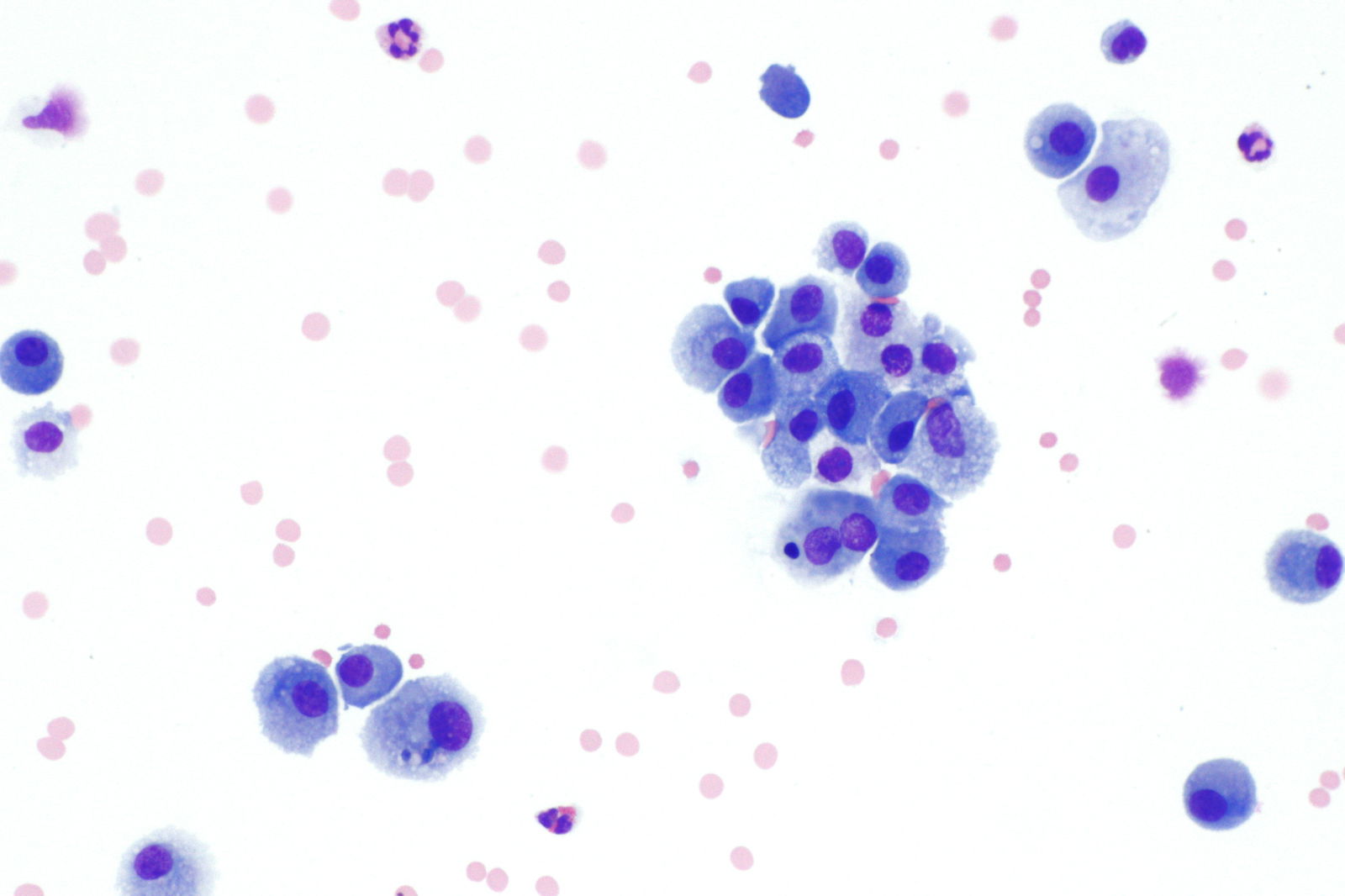
Fig. 8. Macrophages (a type of white blood cell). Macrophages accumulate in large numbers and arrive after the initial neutrophil response to clean up dead neutrophils, cellular debris, and remaining pathogens. Image by Librepath via Wikimedia Commons.
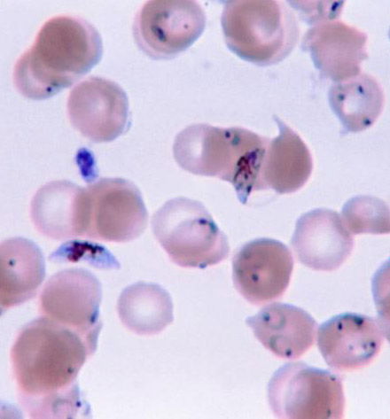
Fig. 9. Plasmodium falciparum, the causative agent of malignant malaria. Notice the trophozoite rings inside the red blood cell. Image by Filip em via Wikimedia Commons.
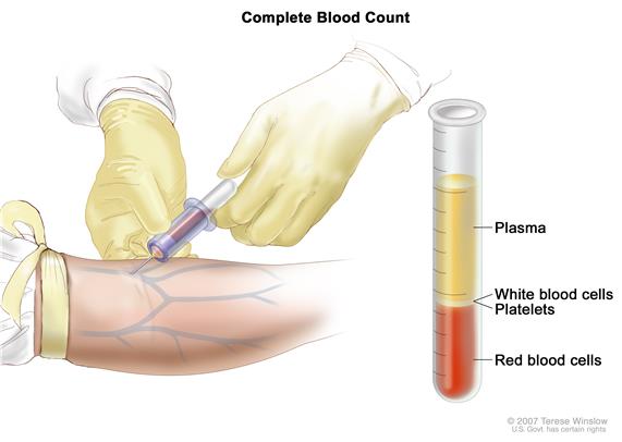
Fig. 10. Complete Blood Cell Count is taken from clinical sample. Image from National Cancer Institute.
For example, a high macrophage count would indicate a bacterial infection (Fig. 8). If you eat food contaminated with E. coli, then your neutrophil count will go up in the first week, and then the macrophage count will follow to combat the infection. If you come down with a malaria infection, then the parasite (Plasmodium) will form rings in the RBCs (Fig. 9). In the specific case of malignant malaria Plasmodium falciparum, three trophozoite rings will form, and later hemozoin (black dots of metabolic waste) will form in erythrocytes (Gillen and Sherwin 2013). Blood has the power to reveal a diagnosis, and a doctor can prescribe proper treatment (Fig. 10).
Blood has the power to reveal a diagnosis and a doctor can prescribe proper treatment (Fig. 10).
Summary and Conclusions
In blood, we study the reason for our existence.
The largest contributors to the overall number of human cells are RBCs. Almost 90% of human body cells (estimated) consists of RBCs, WBCs, and platelets, while the other 10% consists of nucleated cells. The striking dominance of the hematopoietic lineage in the cell count (90% of the total) is counterintuitive given the composition of the body by mass. Blood reveals an Intelligent Designer, a Master Craftsman. In blood, we study the reason for our existence. Blood is one substance that the human body depends upon for life. William Harvey and Anton van Leeuwenhoek not only revolutionized our understanding of the heart and circulation, but they also illuminated our knowledge of blood. Harvey said: “Blood is the cause not only of life in general but also of longer or short life, of sleep and watching, of genius, aptitude, and strength. It is the first to live and the last to die” (Brand and Yancey 1984, 66).
Blood signifies life to the medical practitioner. It feeds every cell in the body with its precious nutrients. When it slips away, our body falters. If we know Christ, then, we will be given a new body (perhaps with perfect blood) in heaven. Our bodies may not be perfect this side of heaven; however, we can praise God that our bodies are made fearfully and wonderfully. Science confirms that life is in the blood!
References
Behe, Michael J. 1996. Darwin’s Black Box: The Biochemical Challenge to Evolution. New York: The Free Press.
Brand, P. and P. Yancey. 1984. In His Image. Grand Rapids, Michigan: Zondervan Publishing Co.
Gillen, A. L., F. Sherwin, and A. C. Knowles. 2001. The Human Body: An Intelligent Design, 2nd ed. St. Joseph, Missouri: Creation Research Society Books.
Gillen, A. L., and F. Sherwin 2013. The Genesis of Malaria. The Origin of Mosquitoes and Their Protistan Cargo, Plasmodium falciparum. Answers in Depth 8 (June 19, 2013). https://answersingenesis.org/biology/disease/the-genesis-of-malaria/.
Gillen, A. L. 2009. Body by Design: Fearfully and Wonderfully Made, 6th printing. Green Forest, Arkansas: Master Books.
Gillen, A. L., and J. Conrad. 2014. Our Impressive Immune System: More than a Defense. Answers in Depth 9 (January, 15, 2014). https://answersingenesis.org/human-body/our-impressive-immune-system-more-than-a-defense/.
Gillen, A. L. 2012. Antony van Leeuwenhoek: Creation “Magnified” through His Magnificent Microscopes. Answers in Depth 7 (August 15, 2012). https://answersingenesis.org/creation-scientists/profiles/antony-van-leeuwenhoeks-microscopes-creation-magnified/.
Gillen, A. L., and J. D. Oliver. 2009. Creation and the Germ Theory: How a Biblical Worldview Helped Shape the View that Germs Make Us Sick. Answers in Depth 4 (July 29, 2009). https://answersingenesis.org/biology/microbiology/creation-and-the-germ-theory/.
Hoole, S. 1798. The Select Works of Antony van Leeuwenhoek, containing his Miscrosopical Discoveries in many of the Works of Nature. 2 vols. London: G. Sidney, 1798; New York: ECCO Press, 2012 reprint.
Marieb, E. N. and Hoehn, K. 2020. Anatomy and Physiology, 7th edition. Pearson Benjamin/Cummings Pub. Co. San Francisco.
Sender, R., S. Fuchs, and R. Milo. 2016. Revised Estimates for the Number of Human and Bacteria Cells in the Body. PLoS Biology, 14(8):e100253. doi:10.1371/journal.pbio.1002533.
Wikipedia. 2019. Complete Blood Cell definition. https://en.wikipedia.org/wiki/Complete_blood_count.
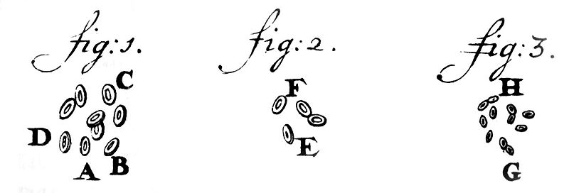
Red Blood Cells drawn by Leeuwenhoek in 1719. Image by Deitzel65 via Wikimedida Commons.
More Quotes and Notes
RBCs live for about 120 days, at which point they are removed from circulation by macrophages in the spleen and liver. At the same time, specialized stem cells are replacing the dead RBCs at roughly the same rate.
my work, which I’ve done for a long time, was not pursued in order to gain the praise I now enjoy, but chiefly from a craving after knowledge, which I notice resides in me more than in most other men. And therewithal, whenever I found out anything remarkable, I have thought it my duty to put down my discovery on paper, so that all ingenious people might be informed thereof.
–Antony van Leeuwenhoek, letter of June 12, 1716
is a Professor of Biology at Liberty University. Coauthor is a graduate student in the Department of Biology & Chemistry at Liberty University.
Answers in Depth
2019 Volume 14

Answers in Depth explores the biblical worldview in addressing modern scientific research, history, current events, popular media, theology, and much more.
Browse Volume
Support the creation/gospel message by donating or getting involved!

Answers in Genesis is an apologetics ministry, dedicated to helping Christians defend their faith and proclaim the good news of Jesus Christ.
- Customer Service 800.778.3390
- © 2024 Answers in Genesis